
Association of a medial UKA and a Patellofemoral Arthroplasty: is it possible?
317
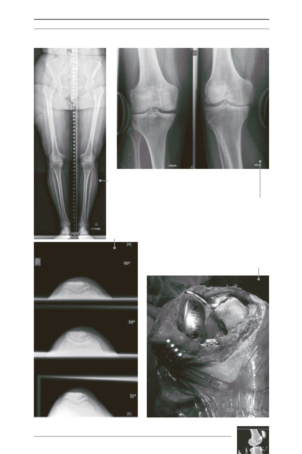
Fig. 1: This full-length X-ray is showing a medial arthritis of
the knee on a very-active 61 years-old woman. She presented
with a medial and patell-femoral pain without any lateral
pain, a stable knee and failure of the medical treatment.
Fig. 2: The varus and valgus stress X-rays are very important to
control the reducibility of the deformity and to check the full
thickness of the cartilage on the non-resurfaced compartment.
Fig. 3: Patello-femoral sky views are important to
assess the wear on the patella-femoral compartment
and the position of the patella.
Fig. 4: This intra-operative view of the knee opened
through a sub-vastus approach is showing the trial
implants in place on the patella-femoral and the medial
compartment with preserved cruciate ligament.


