
S. Parratte, M. Ollivier, J.M.Aubaniac, J.N. Argenson
318
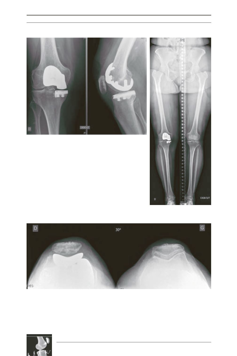
Fig. 5: The 1-year post-operative X-rays are showing a correct implant
positioning on the ML and on the AP views.
Fig. 7: Patello-femoral sky view at 30° is showing a proper position of the trochlear implant below the
patella.
Fig. 6: The postoperative full-length X-ray is showing a proper
restoration of axis of the lower limb without any over correction of
the deformity.


