
Acute Patellar Dislocation – Mini- Battle- Conservative treatment versus surgical treatment
65
The Kujala score was 69 in the conservatively
treated group and 92 in the surgically treated
group.
Our results from conservative treatment greatly
resembled those of Maenpaa and Letto [3],
who found that 44% of their patients presented
recurrent dislocation over a 13-year follow-up
period.
In analyzing this series, we found that the
conservative treatment did not follow any
pattern. The patients’ adherence to use of
immobilization andphysiotherapywas variable.
The patients who were treated surgically
presented worse results when their deinsertions
occurred in the ligament substance and in the
femoral insertion. This result is in agreement
with the observations of Sillanpaa
et al.
[7], in
a study on deinsertions of the MPFL in the
femur, who also considered that these had a
worse prognosis.
The better results from surgical treatment than
from conservative treatment, with absence of
recurrent dislocation, were in agreement with
those of Nomura
et al.
[8], Ahmad
et al.
[9] and
Sallay
et al.
[10].
Randomized study
We organized a second series, now in the form
of a randomized study, in which one group of
orthopedists had the assignment of guiding and
following up conservative treatment and other
group, surgical treatment. Radiography and
MRI were performed on all the patients, who
were then divided into two groups [11].
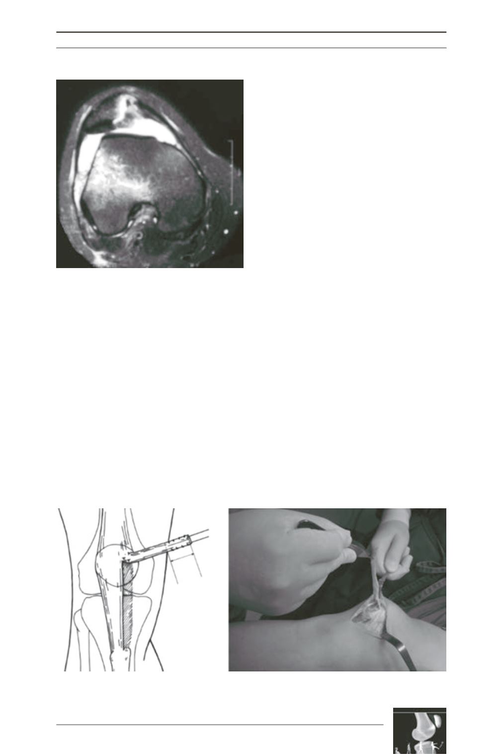
The surgical treatment consisted of a MPFL
reconstruction technique using a 0.5cm strip
from the medial patellar tendon, which was
kept inserted in the proximal third of the patella
(fig. 3) and was fixed in the region between the
medial femoral epicondyle and the tubercle of
Fig. 2 : Magnetic resonance imaging demonstrating
patellar dislocation with signs of bruising of the
lateral femoral condyle and fracturing due to
tearing of the medial edge of the patella.
Fig. 3 : Harvesting of 0.5cm graft from the patellar ligament after its deinsertion from the tibia.


