
W. Fitz
130
Table 1: Medial and lateral femoral Ap and ML dimensions are different
and there are significant differences between males and females [3]
Total
Males
Females
p-Value
Comparing
Males
To females
Height (cm)
168.7 (10.3)
173.6 (9.1)
163.4 (8.9)
p < 0.001
Weight (kg)
73.8 (21.9)
74.9 (22.2)
72 ?6 (20.6)
p = 0.715
Medial Tibia AP Length
5.06 (0.46)
5.37 (0.38)
4.75 (0.29)
p < 0.001
Medial Tibia ML Width
3.04 (0.32)
3.27 (0.25)
2.82 (0.19)
p < 0.001
Lateral Tibia AP Length
4.74 (0.46)
5.03 (0.34)
4.45 (0.38)
p < 0.001
Lateral Tibia MpL Width
3.21 (0.32)
3 .41 (0.26)
3.01 (0.23)
p < 0.001
Medial Condyle AP Length
5.73 (0.45)
6.01 (0.33)
5.45 (0.37)
p < 0.001
Medial Condylar ML Width
2.61 (0.29)
2.80 (0.23)
2.43 (0.22)
p < 0.001
Lateral Condyle AP Length
6.23 (0.51)
6.55 (0.35)
5.92 (0.45)
p < 0.001
Lateral Condylar ML Width
2.85 (0.33)
3.09 (0.25)
2.61 (0.19)
p < 0.001
Med/Fem Art. Surface AP Length 4.84 (0.41)
5.04 (0.35)
4.65 (0.38)
p < 0.001
Lat/Fem Art. Surface AP Length 4.46 (0.47)
4.71 (0.41)
4.22 (0.41)
p < 0.001
Lateral Tibia AP/ML Ratio
1.48 (0.09)
1.48 (0.11)
1.48 (0.08)
p = 0.869
Medial Tibia AP/ML Ratio
1.67 (0.09)
1.64 (0.10)
1.69 (0.08)
p = 0.093
Lateral Condyle AP/ML Ratio
2.20 (0.17)
2.13 (0.17)
2.27 (0.12)
p = 0.002
Medial Condyle AP/ML Ratio
2.21 (0.18)
2.16 (0.19)
2.25 (0.18)
p = 0.080
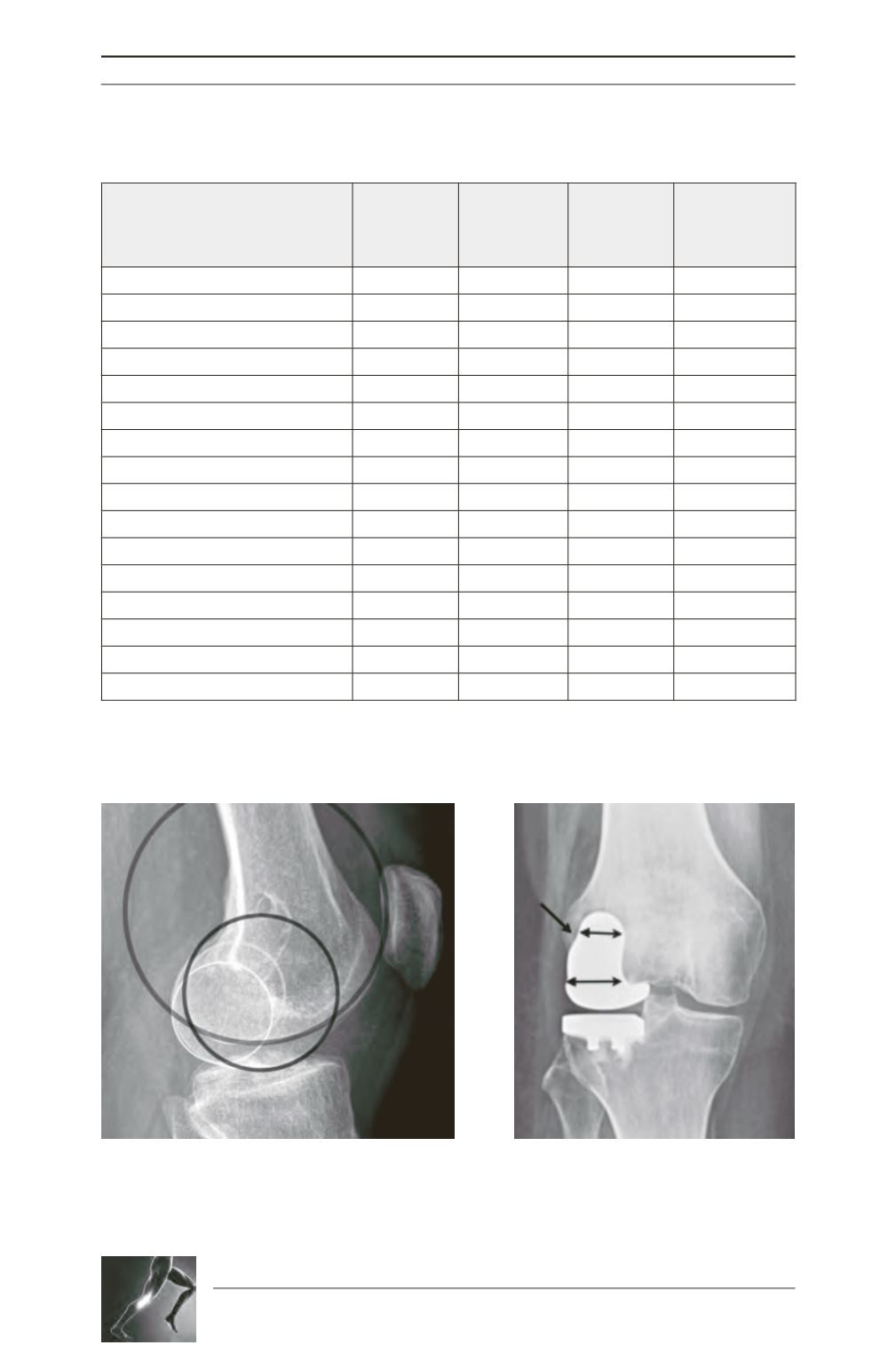
Fig. 1: Medial and lateral femoral condyles have similar
posterior but different anterior radii [1]. The anterior
lateral radius is twice as large compared to the
anterior medial radius.
Fig. 2: Different geometry of the lateral
femoral condyle showing a custom lateral
UKA. Theposterior condyle ismuchnarrower
compared to the width more anteriorly.


