
Physeal-sparing MPFL reconstruction in children
49
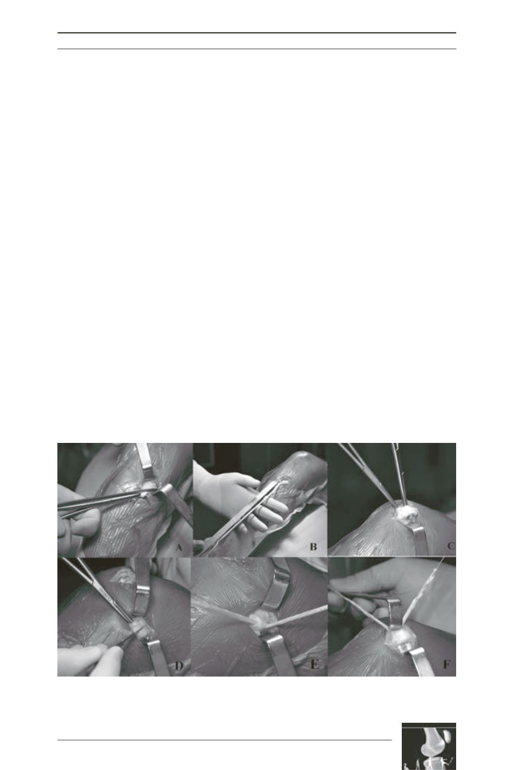
collateral ligament as a pulley as described by
Deie [2] (fig. 2E). The three different layers of
the medial patellar system were dissected: the
first corresponds to the superficial retinaculum,
the second to the MPFL and MCL and the third
to the knee capsule. The tendon bundles were
transferred from the pulley to the patella
locating in the second layer taking care they
were not twisting each other.
Through the patella approach, two transversal
bone tunnels are drilled with the size of the
semitendinosus tendon (fig. 2C). Each graft
bundle is passed through the patella tunnels
[1]. After the graft is passed in the tunnels
(fig. 2F), it is passed back and sutured to itself
pulling the patella medially. Tensionning the
graft is performed with the knee in 30° of
flexion. The correct among of tension prevents
lateral subluxation without causing medial
subluxation or excessive medial compression.
Patella tracking and stability are tested throught
the range of knee motion. In full extension, the
patella slightly moves medially (favorable non
isometry).
Selected associated procedures are performed
during the same time
(Cf. global strategy)
.
The tourniquet is deflated and after accurate
hemostasis, an intra-articular drainage is or no
associated. The wounds are closed with
interrupted
subcutaneous
sutures
and
absorbable running subcuticular sutures. Elastic
bandagewrapandpostoperative immobilization
amovible brace with 10° flexion are used.
Postoperative care: full weight-bearing is
allowed immediately. Early active range of
motion exercices are started as tolerated.
Immediate continuous passive motion is
associated. A return to sport and full activities
is allowed after 4 to 6 months.
Method
Results were assessing with Kujala scoring
system. Objective correction of the patella tilt
was measured with CT scan preoperatively and
postoperatively 1 year following surgery.
Fig. 2: Details and intraoperative views of different steps for MPFL reconstruction in our unit.


