
F. Chotel, A. Peltier, A. Viste, M.M. Chaker, J. Bérard
48
TAGT distance > to 20mm [6, 8]. In such
situation and for adolescents with closed
growth plate, a classical tibial tubercule
osteotomy is used. More rarely, and for
adolescents a trochleoplasty according to
Dejour is associated. In younger children, and
whendealingwithpermanent patelladislocation
which combine short quadriceps, a Judet
quadriceps liberation is associated.
Surgical procedures
Since 2007
, we perform MPFL reconstruction
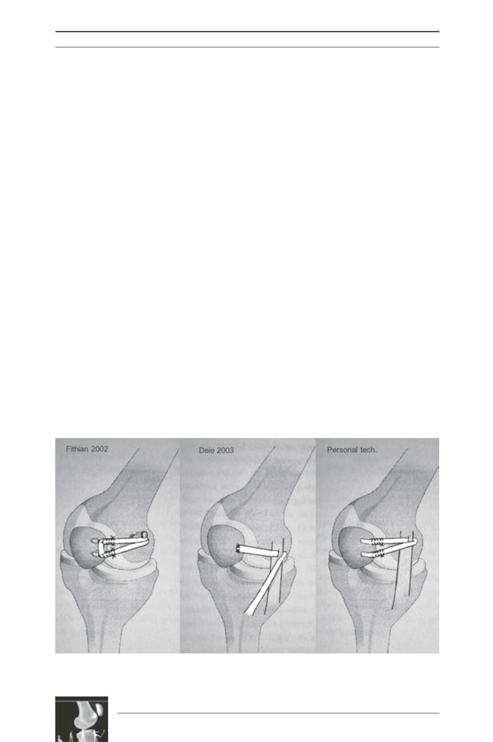
in children. Initially we used Deie technique
for skeletally immature patients [2] and Fithian
technique for adolescents [1] (fig. 1). This
preliminary experience about 13 cases has been
reported during EPOS meeting in 2010 [3]
(Results).
In the mid-2010, we develop an anatomic
double bundle physeal-sparing technique using
a free semitendinosus tendon
. This technique
will be reported here (fig. 2).
This technique is mixing Fithian method and a
modified Deie method (fig. 1). It is conducted
under general anaesthesia and continuous loco-
regional analgesia. The patient is in supine
position. The operated knee is placed at 80°
flexion with a foot support distally and a
support on the lateral aspect of the thigh allows
stability during surgery.Anon-sterile tourniquet
is placed as proximal as possible.
The procedure starts with semitendinosus
transplant harvest. A longitudinal and oblique
2cm skin incision is made medial to the tibial
tubercule. The semitendinosus is identified,
and its tendon is harvested proximally with a
tendon stripper first (fig. 2A-B), while it distal
insertion is also separated. A running 2-0
resorbable crisscross suture is placed in both
end of the free tendon.
Two additional 2cm longitudinal incisions are
made at the superomedial border of the patella
and over the medial epicondyle. Through the
epicondyle approach, the medial collateral
ligament is dissected on an O’shaughnessy
dissecting forceps (fig. 2D). A space is created
between 2/3 anterior and 1/3 posterior of MCL
proximal fiber. The tendon of semitendinosus
is transferred to the patella using the posterior
one-third of the femoral insertion of the medial
Fig. 1: Illustration of Fithian, Deie and personal technique for MPFL
reconstruction in children and adolescent.


