
Anatomical positioning of the medial patellofemoral ligament in children
43
The aponeurosis of the VMO is sutured back to
the patella using Vicryl, with further closure of
subcutaneous tissues and skin. Routine
dressings and bandages are applied.
Rehabilitation
Postoperatively partial weight-bearing using
crutches was allowed. Daily physiotherapy
with active and passive flexion and extension
exercises of the knee, strengthening of the
vastus medialis muscle and straight leg-raising
exercises were recommended. Full weight-
bearing was allowed at two weeks and return to
sport was allowed at the third postoperative
month.
Discussion
Whereas there are numerous publications about
anatomical reconstruction of the MPFL in
adults to our knowledge this is the first report
about anatomical reconstruction of the MPFL
considering the relation of its femoral insertion
to the distal femoral physis in children.
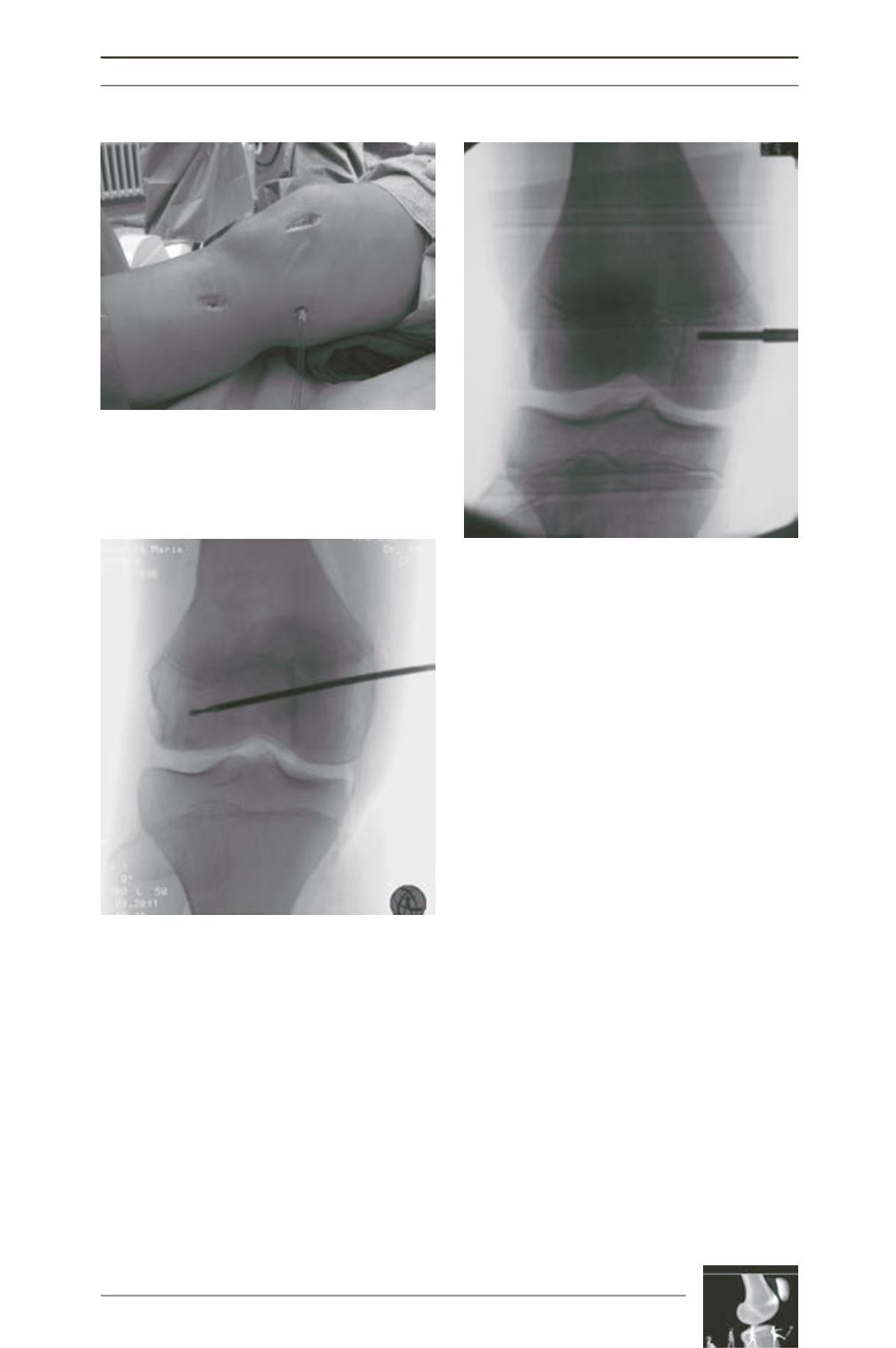
Fig. 3 : The free ends of the graft are pulled between
the second and third layer to the femoral insertion
point.
Fig. 4 : After identification of the entry-point the
guide-pin is drilled to the lateral condyle strictly
distal to the physis.
Fig. 5 : Fixation of the graft within the medial
condyle tunnel using a bioresorbable interference
screw.


