
T. Ait si selmi, C. Murphy, M. Bonnin
188
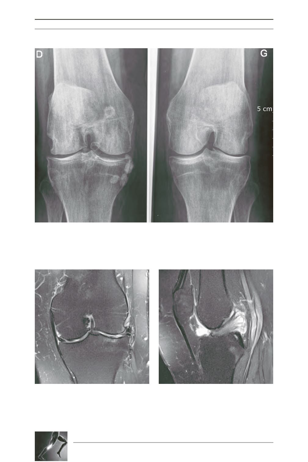
Fig. 8: Lateral OA with a narrowed inter-condylar notch.
Fig. 9: MRI showing ACL fibres engulfed in
osteophytes of the inter-condylar notch (X-Ray on
figure 8).
Fig. 10: MRI showing a degenerative ACL cyst.
Note that the anterior ligament bundle is clearly
seen.


