
S. Zaffagnini, P.G. Ntagiopoulos, D. Dejour, B. Sharma-Dort, S. Bignozzi, N. Lopomo, F. Colle
144
and 2-0 braided sutures (Vicryl) (fig. 3). The
superior 2/3
rd
of the medial border of the patella
was exposed and freshened via sub-periosteal
dissection, without damaging the synovial
membrane. Two antero-posterior tunnels,
3.5mm wide each, were drilled short of the
patellar articular surface, 10mm below superior
patellar pole and 15mm lateral to medial
patellar pole, separated with 20mm bone in
between. These tunnels were connected in a
U-shaped tunnel, while a pulley was fashioned
in the medial retinaculum, 1cm medial to the
patella (fig. 4). After dissection of the MPFL,
the graft tunnel was positioned in the antero-
superior part of the native insertion, using a
7mm drill, over a k-wire, without violating the
lateral femoral cortex (fig. 5). The graft was
passed with suture passers, through the
U-shaped patellar tunnel, under the pulley and
into the blind femoral tunnel, when the knee
was cycled 10 times. The tension in the MPFL
was set to allow one quadrant lateral translation
of the medial facet with a firm end-point, with
out a medial tilt. The tension was maintained,
while the graft was fixed with interference
screws, in 70 degrees of knee flexion to ensure
central position in the trochlea.
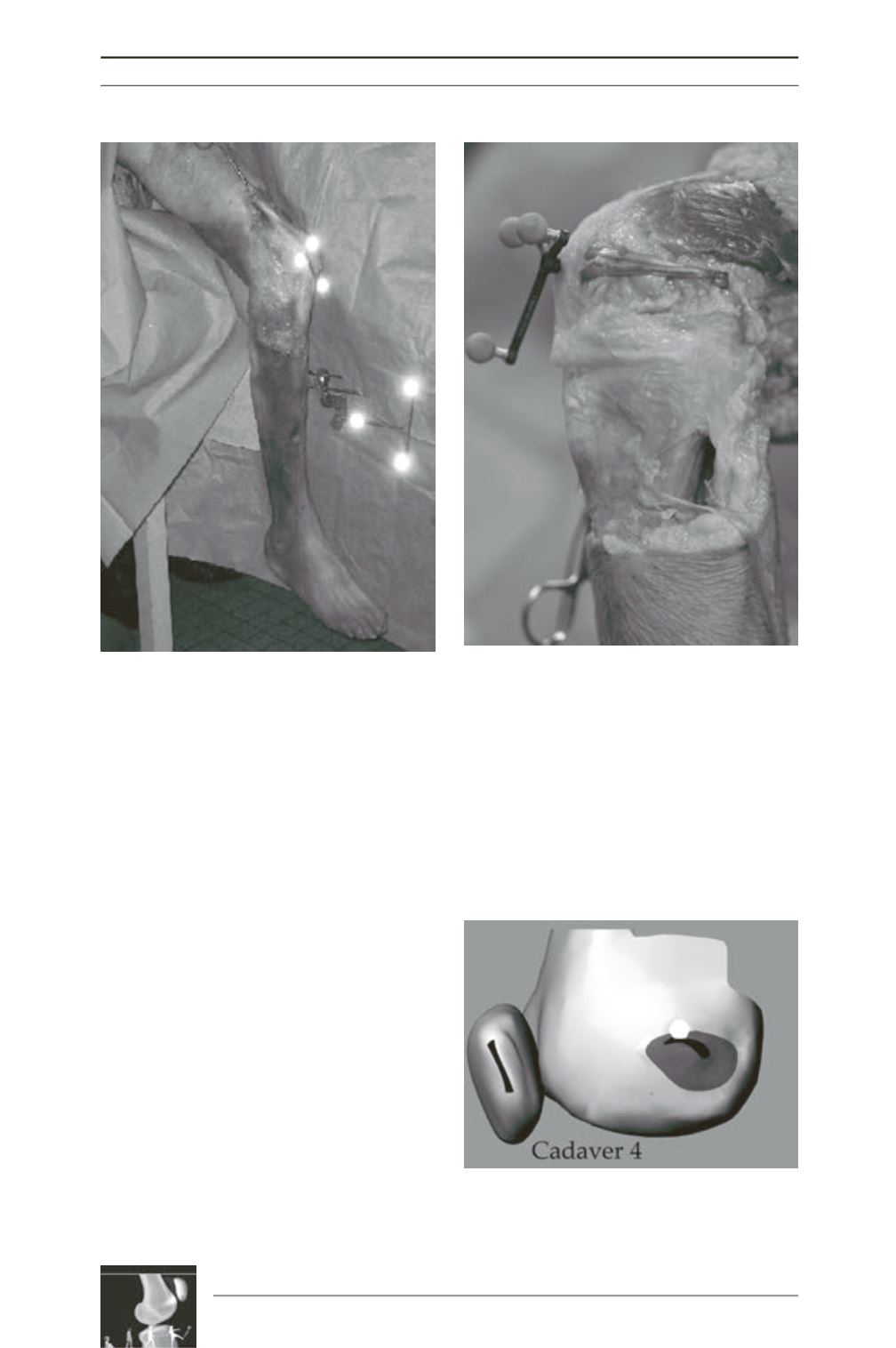
Fig. 2: Experimental Setup showing frames for
navigation and axial quadriceps loading.
Fig. 3: Picture demonstrating a MPFL graft in
position, extending from a single blind femoral
tunnel on the right to a U-tunnel on medial patella
on the left.
Fig. 4: Schematic comparison of Native (black area)
and Graft MPFL Femoral Insertion (white area). The
graft was proximal and anterior to the MPFL
insertion.


