
MPFL Reconstruction: Navigation and Angle of Fixation
145
Statistical Analysis
The mean ± standard deviation of the femoral
insertion of the native MPFL insertion was
compared with that of the graft MPFL. The
native and graft MPFL were compared using
the non-parametric Kruskal-Wallis test in terms
of patellar tracking (p<0.05).
Results
The graft femoral insertion was found to be
proximal and anterior to the geometric center
of the femoral insertion of the native MPFL
(fig. 4). While the difference was statistically
significant in proximal direction, it was not so
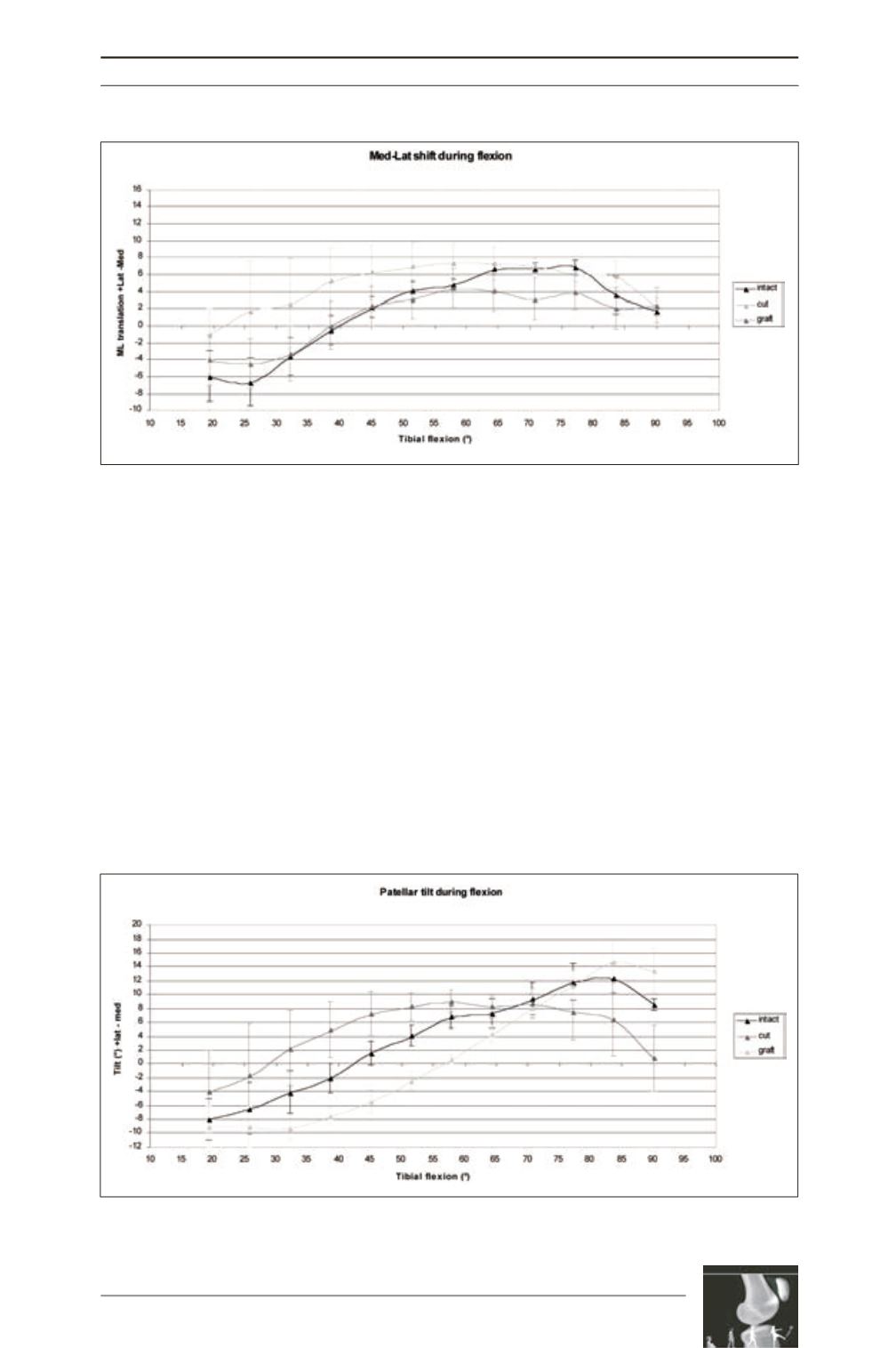
anteriorly. The navigation system recorded a
maximum medial shift in early flexion and
Fig. 5: Medial to lateral patellar translation (shift) in 3 different states of MPFL (intact/cut/reconstructed) in
different degrees of flexion. Medial translation (in mm) expressed in negative (-) values and lateral
translation expressed in positive (+) values.
Fig. 6: Changes of patellar tilt in 3 different states of MPFL (intact/cut/reconstructed) in different degrees
of flexion. Medial tilt expressed in negative (-) values and lateral tilt expressed in positive (+) values.


