


 31 / 242

Place of navigation in 2014: why I resolutely navigate all my TKA?
31
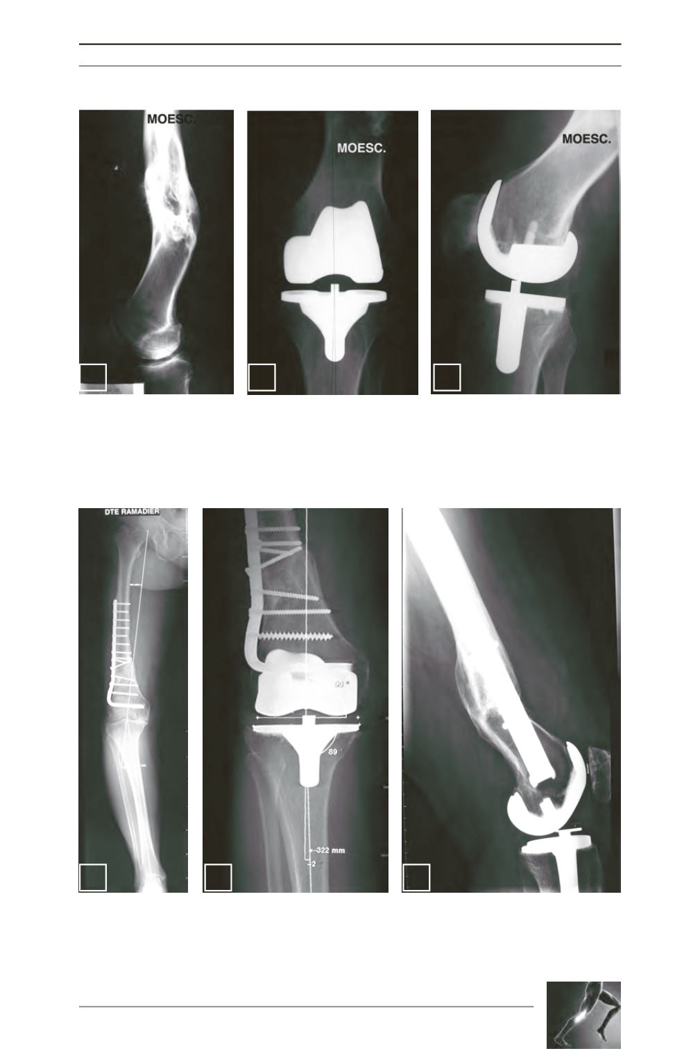
Fig. 7: Lateral view of figure 6.
Fig. 8: Computer-assisted TKA performed in 1999 for figures 6 and 7 case.
Fig. 9: Lateral view of figure 8 case. Notice that the computer placed the femur in flexum to correct the
recurvatum.
Fig. 10: Knee osteoarthritis under a plate, which was implanted 20 years previously. To remove the plate is
not advised because of the risk to lead to recurrent fracture. Moreover, there is also a recurvatummalunion
(see figure 12).
Fig. 11: Computer-assisted TKA of figure 10 case, avoiding to remove the plate.
Fig. 12: Lateral xRay of figure 11 case.
7
8
9
10
11
12