
D. Saragaglia, A. Krayan
30
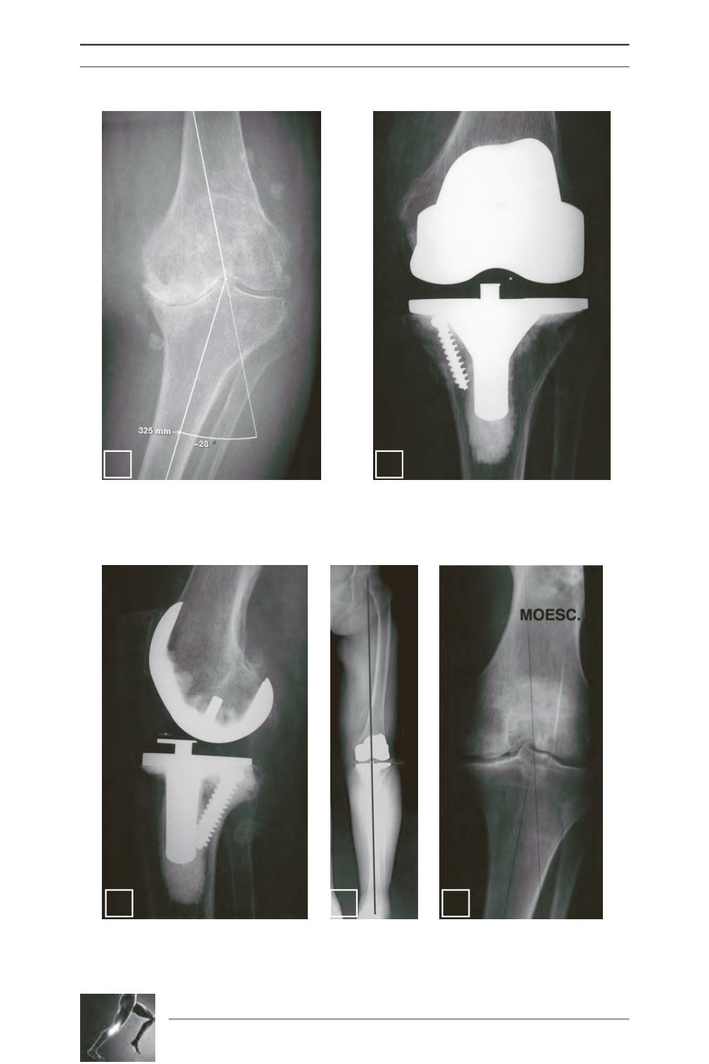
Fig. 2: Severe genu varum deformity with bone loss of the tibial plateau. The varus was reducible
from 28° to 3°!
Fig. 3: Computer-assisted TKA of figure 2 case. A screw was used as a pillar and a mobile bearing
PCL retaining TKA was implanted without any rotation and with only a pie crusting of the medial
collateral ligament.
Fig. 4: Lateral view of figure 3.
Fig. 5: Standing long-leg xRay of figures 3 and 4: HKA at 180°.
Fig. 6: Medial osteoarthritis of the knee occurring on a recurvatum malunion of the distal part of
the femoral shaft.
2
3
4
5
6


