
M. THAUNAT, N. JAN, J.M. FAYARD, C. KAJETANEK, C.G. MURPHY, B. PUPIM, R. GARDON, B. SONNERY-COTTET
130
outside to inside. Next, the suture hook is
passed through the central (the inner portion)
of the medial meniscus. The free end of the
suture in the posteromedial space is grasped
and brought up to posteromedial portal. A
sliding knot (fishing knot type) is applied to the
most posterior part of the meniscus with the
help of a knot pusher and then cut. This
manoeuver is repeated as required depending
on the length of the tear (one knot was inserted
every 5mm for tears limited to the posterior
segment (“limited tears”) (fig. 3a)). Care is
taken during this technique to avoid tangling
the sutures. Once the posteromedial tying is
finished, the knee is positioned in valgus, near
extension and the suture is tested and repeated
if necessary. For some patients, the tear extends
to the midportion of the meniscus and requires
an additional repair through standard anterior
portal with meniscal suture anchor and/or an
outside-in suture (“extended tear”). The
posterior suture is then completed with a repair
through standard anterior portal with a meniscal
suture anchor (Fas T Fix device, Smith &
Nephew, Andover, MA) when the tear extends
to the pars intermedia and/or by Outside-In
sutures with PDS 1 (Ethicon, Inc., Somerville,
NJ) if the tear extends to the anterior segment
of the meniscus (fig. 3b). The stability of the
suture is then tested with the probe.
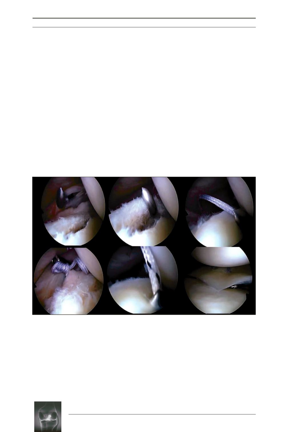
Fig. 2:
Suture of the posterior segment of the medial meniscus of the right knee through a posteromedial
portal with a suture hook device (25° suture lasso loaded with a N°2 fiberstick) (
a, b
) The sharp tip
penetrated the peripheral wall of the medial meniscus from outside to inside. (
c
) Next, the suture hook is
passed through the center (the inner portion) of the medial meniscus. (
d
) The first knot is tied with a knot
pusher. (
e
) A second suture is performed 5mm more posterior to the first one with a tigerstick. (f) The final
suture with non-absorbable suture from the anterior portal.
a
d
b
e
c
f


