
M. THAUNAT, N. JAN, J.M. FAYARD, C. KAJETANEK, C.G. MURPHY, B. PUPIM, R. GARDON, B. SONNERY-COTTET
128
criteria for this study were longitudinal medial
meniscal tears of the peripheral third
(capsulomeniscal junctionor red/red zone) or
junction of the peripheral third with the middle
third (red/white). Complete and partial tears
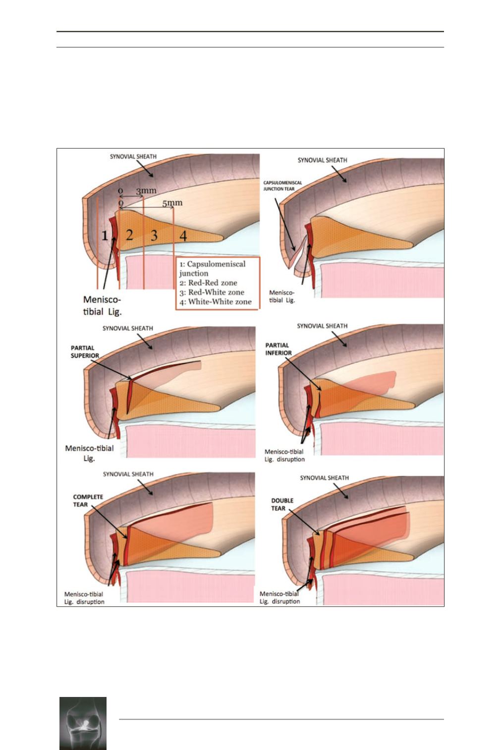
were included (fig. 1). Exclusion criteria were
Fig. 1:
Tear patterns of ramp lesions of the medial meniscus : (
a
) These tears can then further classified by
their proximity to meniscus blood supply, namely whether they are located in the capsulomeniscal junction
1) “red-red” 2), “red-white” 3), or “white-white” 4) zones. (
b
): Type 1: Meniscocapsular junction tear. Very
peripherally located in the synovial sheath. Mobility at probing is very low. (
c
): Type 2: Partial superior lesions.
It is stable and can be diagnosed only by trans-notch approach. Mobility at probing is low. (
d
): Type 3: Partial
inferior or hidden lesions. It is not visible with the trans-notch approach, but it may be suspected in case of
mobility at probing, which is high because of the disruption of the menisco-tibial ligament. (
e
): Type 4:
Complete tear in the red-red zone. Mobility at probing is very high (
f
): Type 5: Complete tears.
a
c
e
b
d
f
Type 1
Type 2
Type 3
Type 5
Type 4


