
C.H. BROWN
144
STRATEGIES TO FIND THE
CENTER OF THE ACL
FEMORALATTACHMENT
SITE
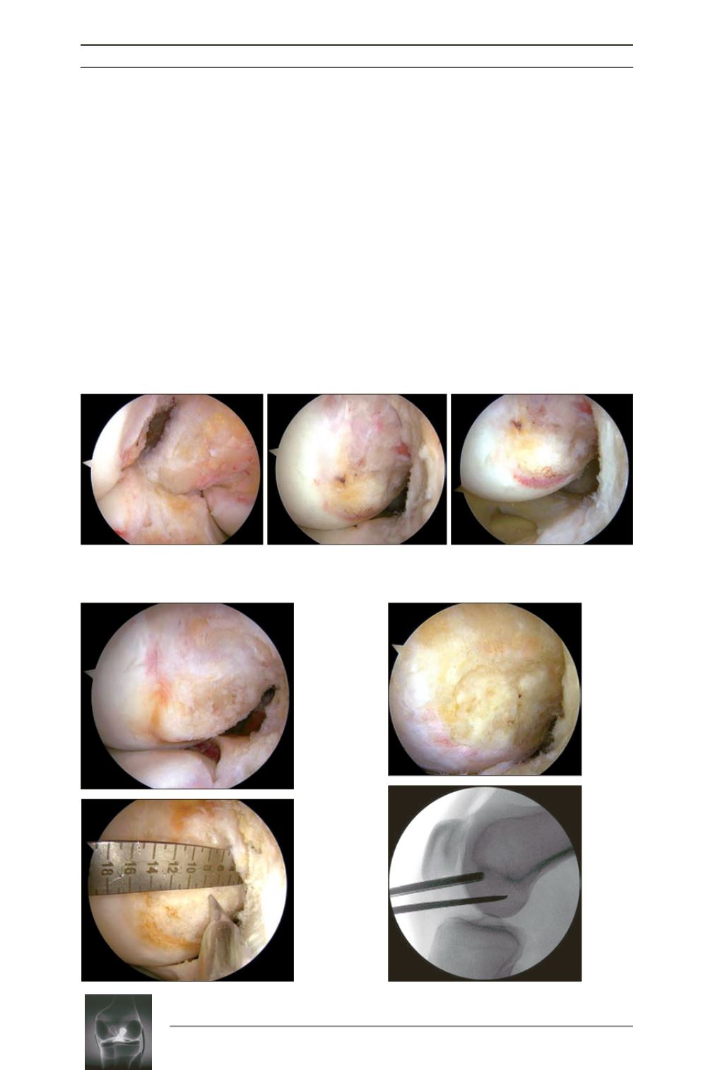
• View the lateral wall of the notch through the
AM portal;
• Place the knee in the figure-four position.
This position opens up the lateral compartment
by lifting the lateral femoral condyle away
from the lateral meniscus, allowing better
visualization of the deep (proximal) part of
the ACL femoral attachment site and the low
(posterior) part of the attachment site where
the indirect ACL fibers (fan-like extension
fibers) insert (fig. 9).
• Use the native ACL footprint if present
(fig. 10).
• Bony ACL ridges (fig. 11).
• ACL ruler (fig. 12).
• Fluoroscopy (most reproducible and accurate
method) ruler (fig. 13).
Fig. 9
Fig. 10
Fig. 11
Fig. 12
Fig. 13
AL Portal View
AM Portal View
Figure-Four: AM Portal View.


