
C. BATAILLER, S. LUSTIG, D. WASCHER, E. SERVIEN, P. NEYRET
14
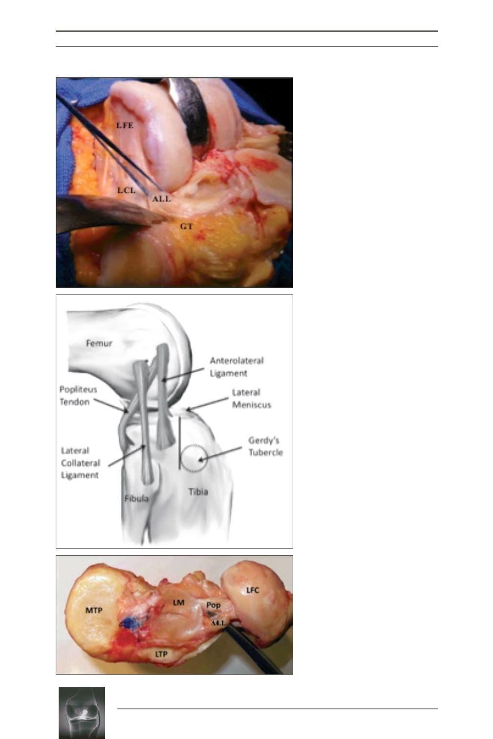
Fig. 1:
Per-operative view during right
knee arthroplasty (a), line drawing of
a right knee (b) and cadaveric
dissection of left knee (c) showing the
ALL described by Vincent
(Photo a:
courtesy of P. Neyret. Photo c and line
drawing from Vincent and al. [5]).
ALL, anterolateral ligament; LCL, lateral
collateral ligament; LFE, lateral femoral
epicondyle; PT or Pop, popliteus tendon; GT,
Gerdy’s tubercle; LM, lateral meniscus; LFC,
lateral femoral condyle.
a
b
c


