
ANTEROLATERAL LIGAMENT HISTORY AND SURGICAL TECHNIQUES
17
which is passed superficial to FCL. The graft is
fixed, with an interference screw, in a blind
tunnel on the LFE [13].
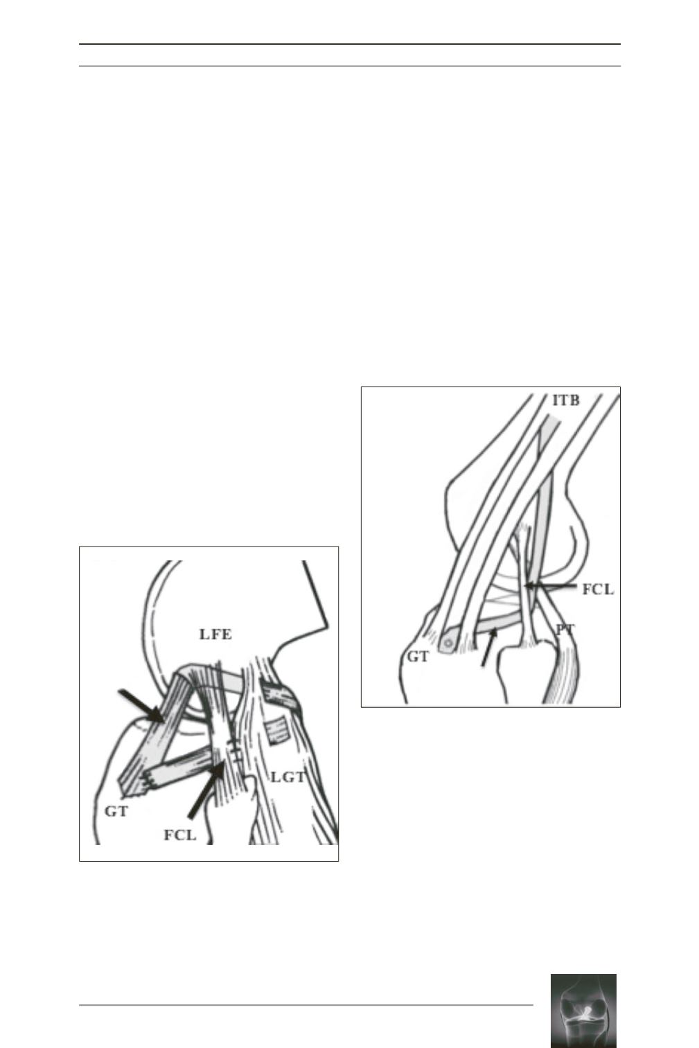
Losee technique: “sling and reef”
operation
[14]
A strip of ITB (16cm long) is harvested and left
attached to the Gerdy’s tubercle. A tunnel was
made through the lateral femoral condyle,
anterior and distal to the attachment of the
FCL. This femoral insertion site closely
approximates the anatomic attachment. The
graft is passed through this tunnel. Then it
passed back through the lateral gastrocnemius
tendon, exiting through the posterolateral
capsule posterior to the FCL and passed under
the FCL. The gastrocnemius tendon,
posterolateral capsule and the graft are all
sutured to the FCL at 45° of knee flexion. Then,
the graft is sutured back to Gerdy’s tubercle
(fig. 4).
Ellison technique
[15]
In 1979, Ellison described a bony transfer of
the ITB distal insertion (fig. 5). A bone block of
1,5cm wide is harvested from Gerdy’s tubercle.
A 1.5cm strip of ITB is released from the bone
block. A passage is made under the FCL. The
capsule beneath the FCL is incised vertically
and plicated. A small hole is then made anterior
to Gerdy’s tubercle, just under the lateral
border of the patella tendon. The bony block is
then passed underneath the FCL and into the
bone trough. It is fixed with a staple at 90° of
knee flexion.
James Andrews technique
[16]
This extra-articular tenodesis used a strip of
ITB of 10cm long, left attached on the tibial
distal insertion. Two small drill holes were
made in the distal femur, corresponding to the
Graft
Graft
Fig. 4:
Line drawing demonstrating the completed
graft position in Losee technique [14]
(from Lerat
and al. [26])
.
LFE, lateral femoral epicondyle; GT, Gerdy’s tubercle;
FCL, fibular collateral ligament; LGT, lateral gastrocnemius
tendon.
Fig. 5:
Line drawing demonstrating the completed
graft position in Ellison technique [15]
(from Lerat
and al. [26])
.
LFE, lateral femoral epicondyle; GT, Gerdy’s tubercle;
FCL, fibular collateral ligament; LGT, lateral gastrocnemius
tendon.


