
THE ILIOTIBIAL BAND WITH ITS FEMORAL ATTACHMENTS AT THE KNEE…
57
By attachment of metal “eyelets” through
which sutures were passed attached to strain
gauges, length changes of lateral soft tissue
structures and of soft tissue grafts for various
operative techniques were tested [10].
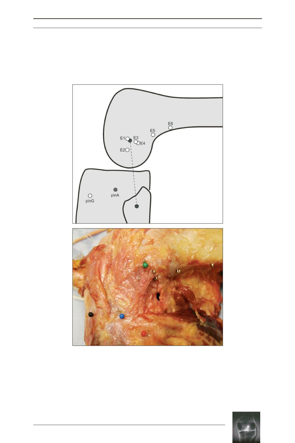
Fig. 5:
Femoral eyelet positioning. Tibiofemoral point combinations
account for structures on the lateral side, extra-articular soft tissue
reconstructions, and femoral isometric points. (
a
) pinG, Gerdy
tubercle; pinA, area of the Segond avulsion; dashed line, lateral
collateral ligament. (
b
)
black
pin, Gerdy tubercle;
blue
pin, area of the
Segond avulsion;
red
pin, fibular head;
green
pin, lateral epicondyle.
(Courtesy of Am J of Sports Med)
.
a
b


