
• Two weeks later, intra-observer (ADK) -
and inter-observer (JH) variation measure-
ments were performed in a subset of
20 patients.
A transparent template at a known diameter of
each squares’ edges of 5 mm was used to avoid
the magnification on the X-ray during the digi-
talization of the conventional stress radio-
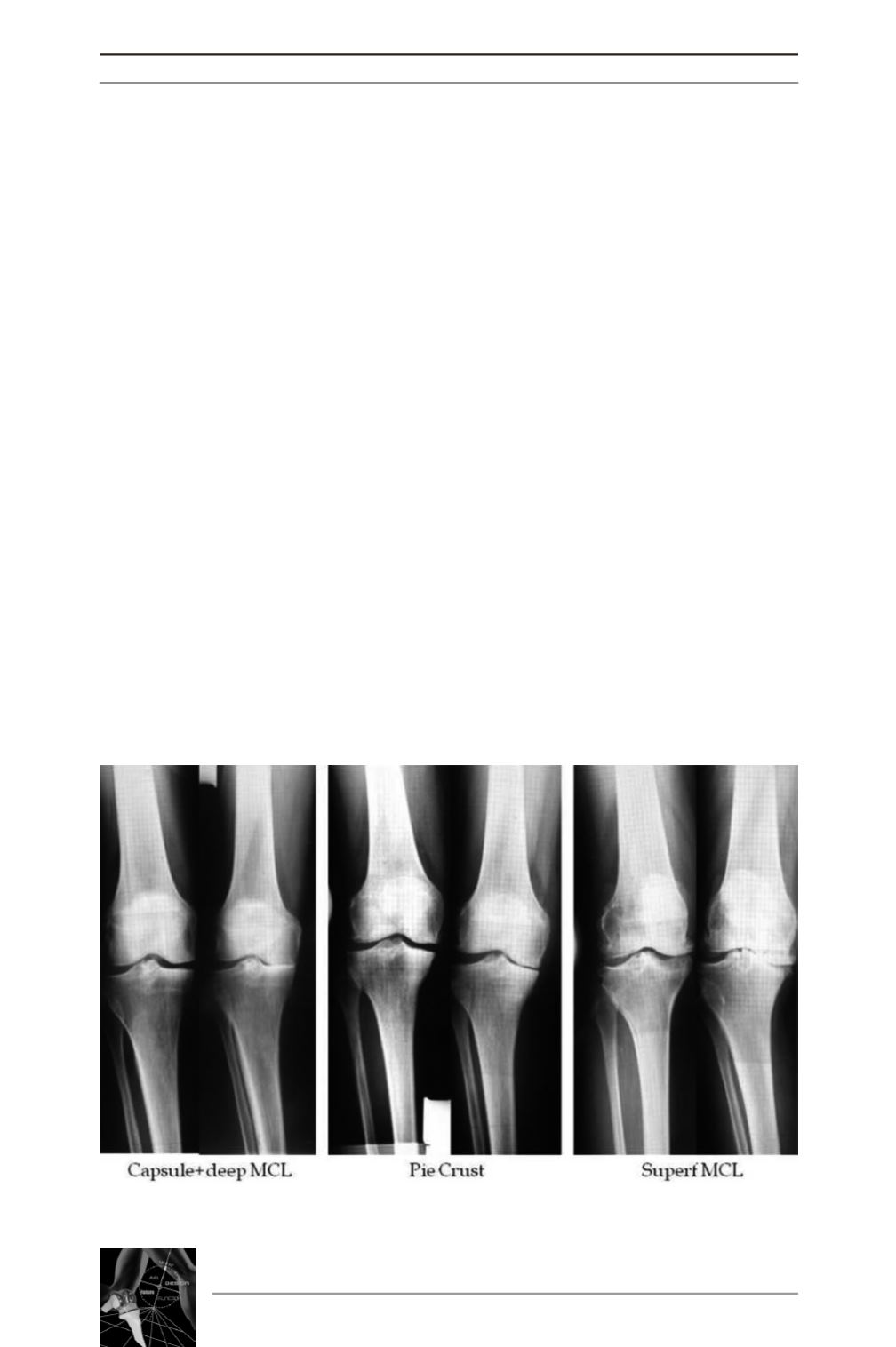
graphs (fig. 5). The digitalized stress radio-
graphs were measured by using National
Institutes of Health ImageJ (version 1.37,
USA). The centers and the lines that were
constructed by one of the observers were not
seen with this software before the measure-
ments were repeated. All the data was stored in
an Excel XP database (Microsoft Corp,
Redmond, WA) and statistical analysis was
carried out by using SPSS for Windows soft-
ware (Version 15; SPSS, Chicago, Illinois).
Intra- and inter-observer correlation was com-
pleted by using Intraclass correlation coeffi-
cient (ICC) method. The Levene’s method was
used to determine the groups’ variance for
homogeneous. Linear regression analysis was
made by Pearson correlation coefficient (r)
test. The differences between the groups were
examined by using One-way Anova test. If
One-way Anova test showed significance,
Bonferroni post hoc test was used for advan-
ced analysis between the groups. The level of
significance was set at P < 0.05.
RESULTS
There was no significant difference of age bet-
ween the groups. All the three groups’ variance
for a.HKA (fig. 6) was homogeneous (p=0.95).
No significant difference was found between
the groups for a.HKA (p=0.08) (Table 1).
The tibiofemoral separation angle
on varus
stress
radiograph showed no significant diffe-
rence (p=0.74) in both groups (Table 1).
Similarly, no significant difference in TFS on
valgus stress was found (p=0.08) between the
groups. As a radiologic parameter, mediolateral
laxity by calculating preoperative TFS did not
differed between the groups to give interest in
the preoperative planning of a medial release.
14
es
JOURNÉES LYONNAISES DE CHIRURGIE DU GENOU
14
Fig. 5 : Radiographic samples of the individuals’ from each group.


