
A philosophy and technique for reconstruction of the medial patellofemoral ligament
153
achieved by pulling the patella proximally with
the bone hook when tying the temporary tape.
When a satisfactory tension pattern, in both the
tape and patellar tendon is achieved, the guide
wire in the epicondyle is overdrilled with a
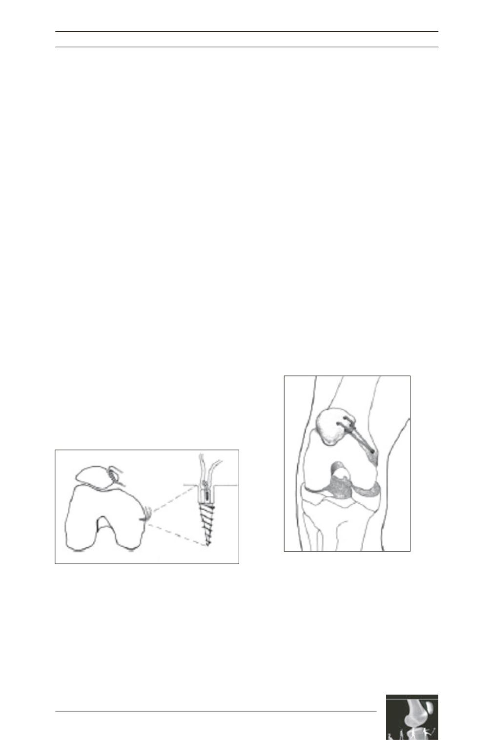
4.5mm cannulated drill. A 5mm bone anchor is
placed in the depth of the drill hole on the
femur. The loop of the double gracilis tendon is
tied into the femoral bone tunnel with the
anchor. The two free ends of the looped tendon
are now brought between the second and third
fascial layers to the exposed medial edge of the
patella and through the two 3mm drill holes on
the medial edge. The free ends of the gracilis
tendon are then folded back on themselves
(fig. 6). The reconstructed ligament is tensed in
the same manner as described above with the
testing tape. Tensing is done with the knee in
full extension while simultaneously pulling
with a bone hook on the patella, in the direction
of the femoral shaft. This manoeuvre prevents
over-tensing of the reconstructed MPFL.
Excessive tension in the reconstructed
ligament can lead to an extensor lag. This
happens when the tension in this reconstructed
ligament is more than in the patellar tendon
with the knee locked in full extension by
maximum quadriceps contraction.
After tensing, the medial and lateral movement
of the operated patella should be similar to that
of the contralateral patella, the idea being to
restore stability to the pre-dislocation situation.
We suggested draping both knees to allow intra-
operative comparison of the patellar movement.
Once the tensing is satisfactory, the free end of
the folded back tendon is sutured to itself and
the surrounding soft tissue with non-absorbable
material (fig. 7). Postoperatively, immediate
full passive motion is encouraged. Active
flexion and light isometric quadriceps exercises
are done. For the first 4 weeks postoperatively
the patient is mobilised partially weight-
bearing, using two crutches. After 4 weeks, the
crutches are discarded and intensive quadriceps
rehabilitation starts. Quadriceps rehabilitation
is often prolonged and can take up to 6 months
or even longer. Normal sports activities can be
resumed as soon as full quadriceps rehabilitation
is achieved.
Fig. 6 : 5mm bone anchor anterior to the medial
epicondyle. Two 3mm drill holes through the medial
patellar rim.
Fig. 7 : Reconstructed MPFL from
the medial epicondyle to the medial
patella.


