
P.J. Erasmus, M. Thaunat
162
the non isometry of the ligament. In unpu
blished cadaveric experiments we found that
that the average length changes in the MPFL
from 0°-90° was 4mm. If the tibial tubercle
was moved 10mm proximally the average
change was 6mm.
When the tubercle was moved 10mm distally
the average length change was only 3mm [9].
Considering this increase in non isometry with
patella alta the distance, at full extension, from
the origin of the MPFL on the medial femoral
condyle to its insertion on the patella, with the
quads fully contracted, is important in planning
surgery. At present there is no specific way to
measure this distance. The most commonly
used measurements for the patella height like
Caton – Deschamps, Blackburne – Peel and
Insall – Salvati measures patella height in
relation to the tibia. What is however more
important is the height of the patella in relation
to the superior border of the trochlea as
suggested by Bernageau [3] on X-rays and
Biedert onMRI’s [4, 1]. Patella alta is associated
with a long patella tendon and patella tendon
length is more sensitive than Caton-Deschamps
index for patella instability [21]?
In reconstructing the ligament the aim should
be to use a ligament that is stronger than the
original to compensate for the underlying
predisposing factors. The reconstructed
ligament should duplicate the non isometry.
The aim of the reconstruction should be to
create a “favourable anisometry” [29] that
duplicates that of the original ligament before
injury. Failure to create favourable non isometry
can lead to redislocation, extensor lag and loss
of flexion. Loss of flexion will also lead to
overload in the patella femoral joint especially
in the medial facet with flexion.
Complications
Loss of motion
In the long term follow up in our series of more
than 200 MPFL reconstructions, done from
1995 till 2008, extensor lag, with full passive
extension and no loss of flexion was the most
common complication. There was no long term
loss of flexion. Not with standing loss of motion
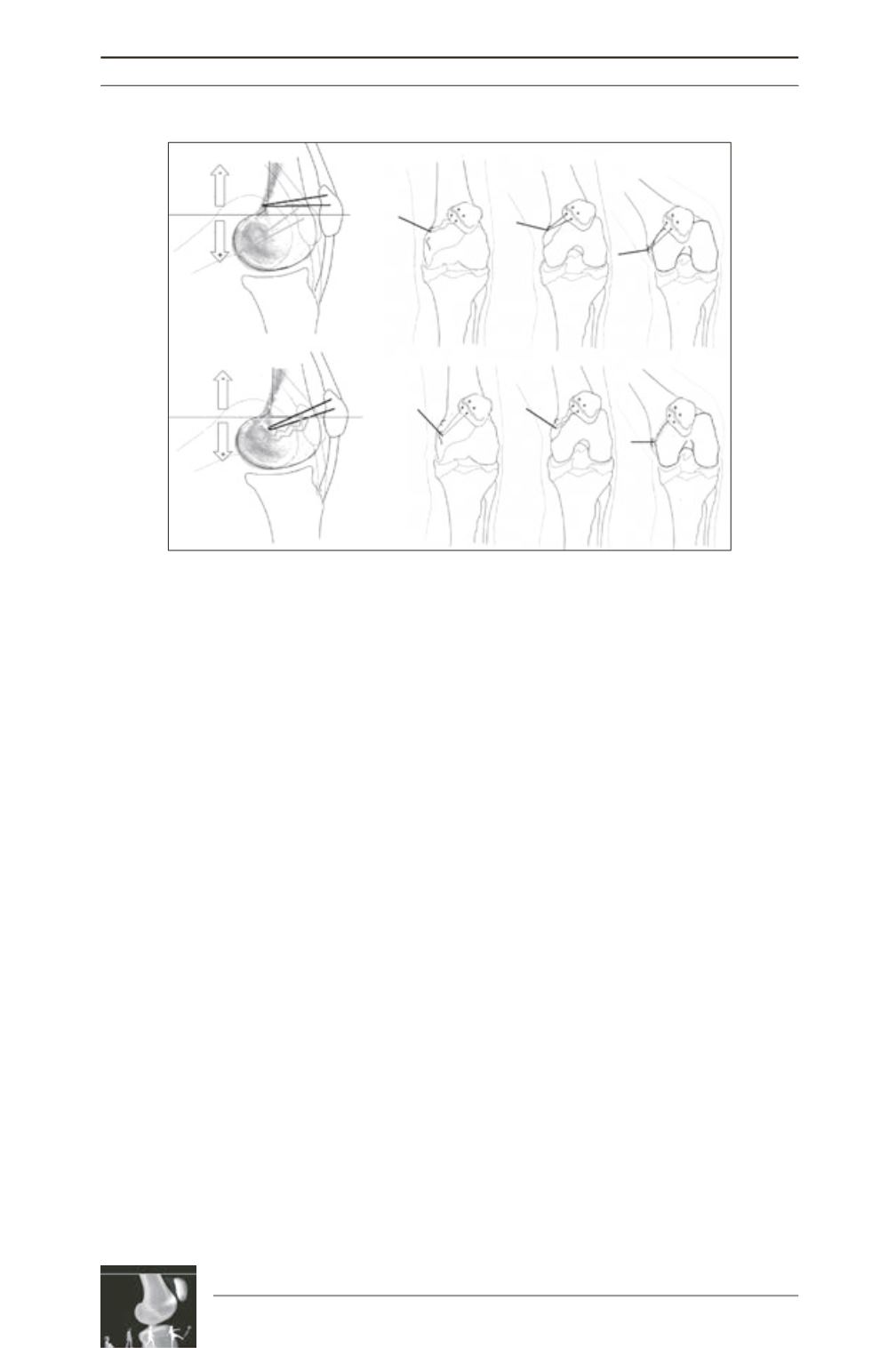
Fig. 1 : A proximal position on the femur will result in a graft that is loose in extension
and tight in flexion, conversely a distal femoral position will result in a graft that is
tight in extension and loose in flexion.


