
Intra- and Interobserver agreement of Dejour´s classification of trochlear dysplasia
183
between low-grade dysplasia, type A from
high-grade dysplasia, type B, C and D was
additionally studied.
Two-grade analysis showed much better results
for intra- and interobserver agreement: The
contingency tables exhibited better results for
intraobserver agreement with proportions of
observed agreement between 56 and 88%
(lateral radiographs) and 70 and 90% (MRI) as
well as for the interobserver agreement with
proportions of observed agreement between 52
and 76% for lateral radiographs and 62 and
96% for MRI-scans.
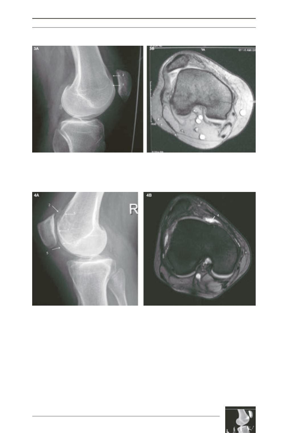
Fig. 3: Example for type B dysplasia.
A: Crossing sign (1), supratrochlear bump (2) on the lateral radiograph.
B: Flat trochlea on the MRI-scan.
Fig. 4: Example for type D dysplasia:
A: Crossing sign (1), supratrochlear bump (2), double contour (3), lateral femoral condyle (5) and the medial
femoral condyle (6) on the lateral radiograph.
B: Asymmetry of trochlear facets plus vertical join (4) on the MRI-scan.


