
P.R.F. Saggin, P. Ferrua, P.G. Ntagiopoulos, D. Dejour
28
knees), although some overhang of values may
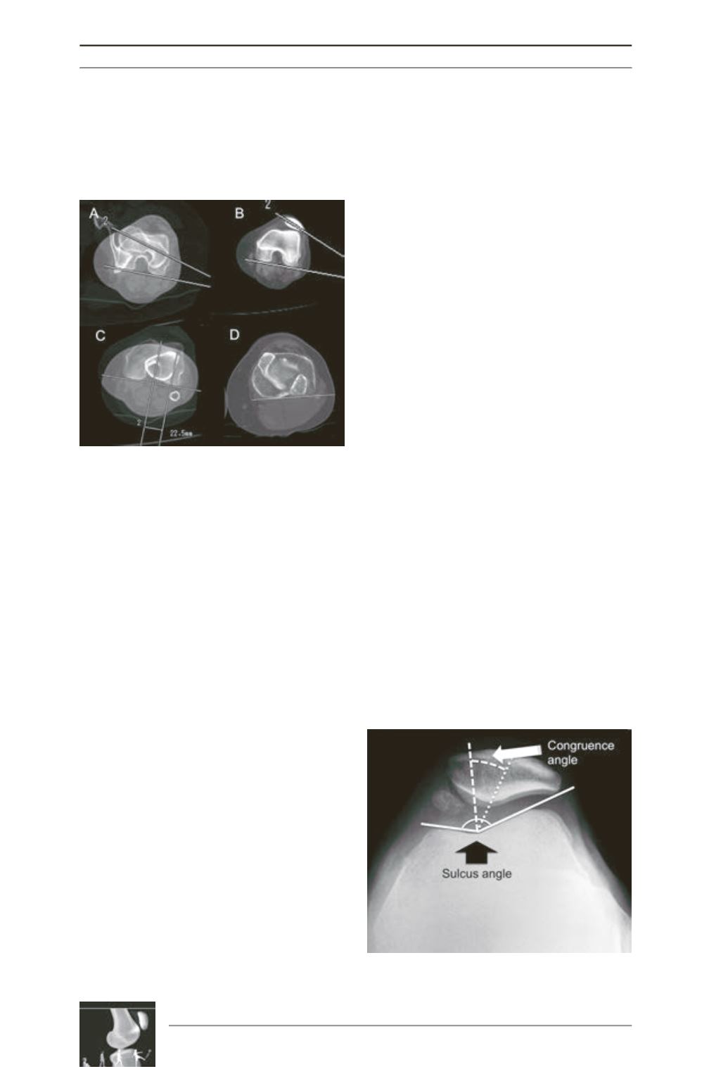
exist. Combined with tilt and TT-TG, these
constitute the Lyon protocol for CT analysis [1]
(fig. 3).
Schoettle
et al.
[21] evaluated the reliability of
the TT-TG on MRI compared to CT scan in
12 knees with patellofemoral instability or
anterior knee pain. The mean TT-TG referenced
on bony landmarks was 14.4±5.4mm on CT
scans, and 13.9±4.5mm on MR images. The
mean TT-TG referenced on cartilaginous
landmarks was 15.3±4.1mm on CT scans, and
13.5±4.6mm on MR images. They found
excellent interperiod (bony vs. cartilaginous
TT-TG), and intermethods (CT vs. MRI
measurement) reliabilities, 91 and 86%
respectively.
Patellar tilt (and
subluxation)
Patellar tilt and subluxation refers to the
abnormal position of the patella in relation to
the trochlear groove. While tilt means increased
lateral inclination of the transverse diameter of
the patella, subluxation refers primarily to
abnormal mediolateral displacement of the
patella in relation to the trochlea. Whether
cause or consequence of instability, they must
be considered for diagnosis and adequate
treatment of instability.
On the lateral view, the shape of the patella is
dependent on its tilt. Normally, the lateral facet
is anterior to the crest. Mild tilt occurs when
both lines (lateral facet and crest) are
superimposed, and severe tilt is when the crest
is anterior to the lateral facet [2].
Methods of evaluating tilt and subluxation have
been described for x-rays axial views:
1)
The congruence angle is measured on
X-rays at 45° of knee flexion. After
measuring the sulcus angle (used to access
trochlear shape), two other lines are drawn
from its vertex: one bisecting the sulcus
angle (reference line) and another to the
apex of the patella. The angle between these
two lines is the congruence angle, considered
positive if the line to the patellar apex is
lateral to the reference line. Average
congruence angle is -6° (SD±11°), and
measures primarily subluxation [6] (fig. 4).
2)
The lateral patellofemoral angle is formed
by one line connecting the highest points of
the medial and lateral facets of the trochlea
and another tangent to the lateral facet of
the patella, drawn on 20° of knee flexion
axial views (Laurin). In normal knees this
angle should open laterally (except in 3% in
which it is parallel). It demonstrates
primarily tilt [22, 23].
Fig. 3: The Lyon Protocol for CT Scan analysis. A:
femoral anteversion; B: external patellar tilt; C:
tibial tubercle-trochlear groove distance (TT-TG);
D: external tibial torsion.
Fig. 4: The sulcus and the congruence angles.


