
25
Imaging in patellofemoral instability must
identify the 4 classic factors implied in the
genesis of the instability – trochlear dysplasia,
patella alta, abnormal tibial tubercle – trochlear
groove distance (TT-TG) and patellar tilt
(excessive patellar tilt with medial ligamentous
disruption) – specially in the chronic setting. In
the acute cases, imaging is sometimes the only
element to provide the diagnosis.
Trochlear dysplasia
Trochlear dysplasia is the most important factor
implied in the genesis of patellar instability [1]
since the femoral sulcus is not sufficient to
provide the osseous restraint to the patella.
Standard lateral X-ray films with perfect
superimposition of the posterior medial and
lateral femoral condyles are the key to the
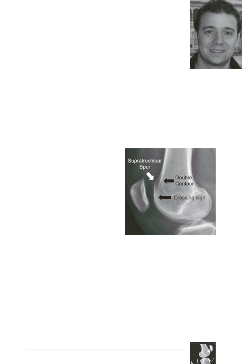
diagnosis of dysplasia. The crossing sign is
typically found in this projection and represents
the point where trochlea becomes flat (the
bottom of the groove reaches the height of the
facets) [1, 2]. Additional findings include the
double-contour sign (representing the
hypoplastic medial facet found posterior to the
lateral one) and the supratrochlear spur (found
in the superolateral aspect of the trochlea) [3,
4] (fig. 1).
Axial X-ray views performed in 45° of knee
flexion allow the measurement of the sulcus
angle [5]. Normal mean value is 138° (SD±6)
[6]. Angles above 150° are found in trochlear
dysplasia. An important issue when analyzing
axial views is that X-rays obtained with higher
flexion angles image the lower part of the
trochlea, frequently missing the dysplasia
present in its upper portion [7]. For this reason,
images obtained at 30° of flexion are
preferred.
Imaging in patellofemoral
instability
P.R.F. Saggin, P. Ferrua,
P.G. Ntagiopoulos, D. Dejour
Fig. 1: Trochlear dysplasia
features on the lateral view.


