
M. Odumenya, S.J. Krikler, A.A. Amis
294
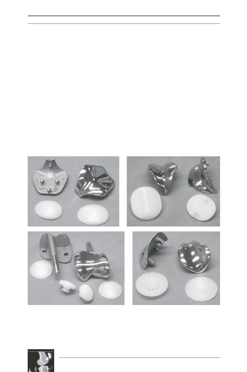
A previous study [14] assessed the pre- and
post-operative tracking kinematics in vitro of
four patellofemoral arthroplasties: Avon,
Blazina, Leicester (Corin Group, Cirencester,
England) and Lubinus (see fig. 5A-D). The
Avon and Leicester implants had tracking paths
that most resembled the native knee.
Unsurprisingly,theBlazina,hadacomparatively
linear pattern following engagement into the
V-shaped trochlear groove. The Lubinus
demonstrated an inconsistent pattern in some
of the specimens. Assessment of these
specimens identified abrupt changes in patellar
tilt indicating the transition point as the patellar
component moved from articulating with the
trochlear component to the native articular
surface.
Patellofemoral joint reaction forces occur as a
result of the tension in the extensor mechanism.
The force vectors in the sagittal plane are
composed of the quadriceps and patellar tendon
tensions (see fig. 6). The result is a force applied
by the patella posteriorly onto the anterodistal
femur. In the coronal plane this force has a
lateral component caused by the Q angle. Due
to the compressive nature of the force, loosening
is unlikely to occur during point loading
however, as the force moves position with
increasing knee flexion, large forces will be
applied closer to the proximal edge of the
patella. Numerous fixations pegs cemented into
the patellar bone will resist potential
displacement of the component.
Fig. 5A-D : Patellofemoral joint prostheses: [A] Avon prosthesis, [B] Blazina prosthesis, [C] Leicester
prosthesis and [D] Lubinus prosthesis.
Permission to use image granted by copyright owners Lippincott Williams & Wilkins. Amis AA, Senavongse W,
Darcy P. Biomechanics of patellofemoral joint prostheses. Clin Orthop 2005; 436: 20-9.
A
B
C
D


