
M. Charles, R. Afra, D.C. Fithian
398
More recently, MR imaging has been applied to
the analysis of patellofemoral instability. The
ability of MR images to clearly represent the
articular cartilage has improved the clinician’s
understanding of severe cartilaginous dysmor
phology that was not brought to light with
previous radiographic studies [15-17]. Recent
articles have compared the reliability of MRI
and CT in evaluating the patellofemoral joint [9,
17-23]. Some studies have demonstrated the
accuracy of MRI to evaluate the patellofemoral
joint. However, these articles have also
highlighted discrepancies between previously
established CT and radiographic cutoffs and the
MRI based evaluation of those same measures.
Currently, there is a limited amount of research
applying MRI imaging to patellofemoral
instability. We have investigated some of the
key morphological differences between normal
knees and those with recurrent patellofemoral
instability [24].
Observations
Patellar Tilt
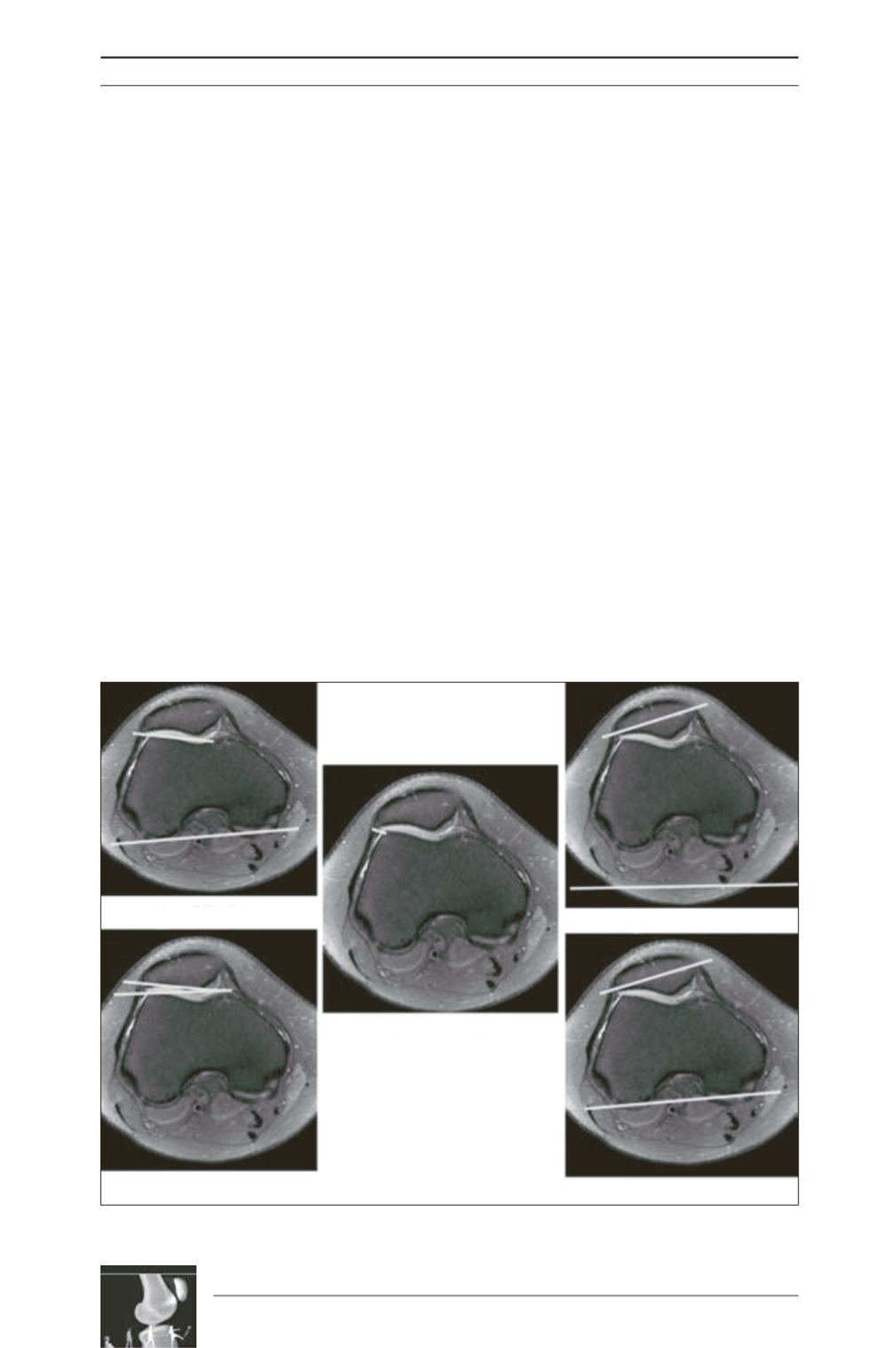
(fig. 1)
As with all the measurements of patellar tilt,
these angles reflect that patients with
patellofemoral instability had an increase in the
lateral rotation of the patella around its superior
to inferior pole. All patellar tilt measurements
were found to be significant between the two
groups.Angle of Laurin (Controls 10.10°±0.48;
PFJDs -5.23°±2.96; p<.001) and Angle of
Fulkerson (Controls 18.18°±0.56; PFJDs
-3.5°±2.62; p<.001) are examples of classic
measures that were found to be significant. The
lateral displacement of the patella (Controls
3.28mm±0.24; PFJDs 6.59±0.69; p<.001) was
also significant.
Fig. 1: Patellar Alignment
Angle of Fulkerson
Angle of Grelsamer
Angle of Laurin
Lateral patella displacement
Patella inclination angle


