
The Clinical Examination in Patellar Instability
95
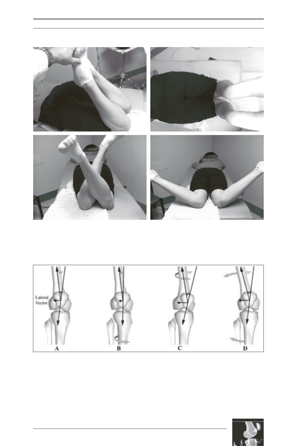
Fig. 2: The prone examination allows assessment of femoral and tibia torsion as well as quadriceps
tightness. (A) evaluation of knee flexion with the hip in extended, which is a measure of quadriceps
flexibility; (B) assessment of tibial torsion; (C) hip external rotation and (D) hip internal rotation.
A
B
D
C
Fig. 3: (A) The Q angle is measured as the angle formed by the intersection of the line drawn from the
anterior superior iliac spine to the midpoint of the patella and a proximal extension of the line drawn from
the tibial tubercle to the midpoint of the patella. Normal alignment of the tibia and femur results in an
offset in the resultant quadriceps force vector (proximal) and the patellar tendon force vector (distal),
creating a lateral vector acting on the patella; (B) tibia internal rotation decreases the Q angle and the
magnitude of the lateral vector acting on the patella; (C) femoral internal rotation increases the Q angle
and the lateral force acting on the patella; (D) knee valgus increases the Q angle and the lateral force
acting on the patella. JOSPT grant to use Figure 1 from Powers CM. The influence of altered lower-
extremity kinematics on patellofemoral joint dysfunction: a theoretical perspective. J Orthop Sports Phys
Ther 2003;33(11):639-646.


