
S. Lustig, C. Scholes, S. Oussedik, M. Coolican, D. Parker
22
The hip was taken through a range of motion
and a digital hip centre generated by the
software package. A midline knee incision was
then performed, prior to a medial parapatellar
arthrotomy. The medial collateral ligament was
elevated from the tibia sufficient to gain access
to the joint. Registration of the epicondyles,
femoral centre, AP axis and femoral condyles
was then undertaken with the navigation
system, followed by registration of tibial
landmarks and malleoli. The lateral epicondyle
was defined as the most lateral prominence of
the lateral femoral condyle, whilst the medial
epicondyle was defined by the medial sulcus of
the medial epicondyle. The navigation software
then generated the surgical trans-epicondylar
axis, a line connecting the 2 epicondyles.
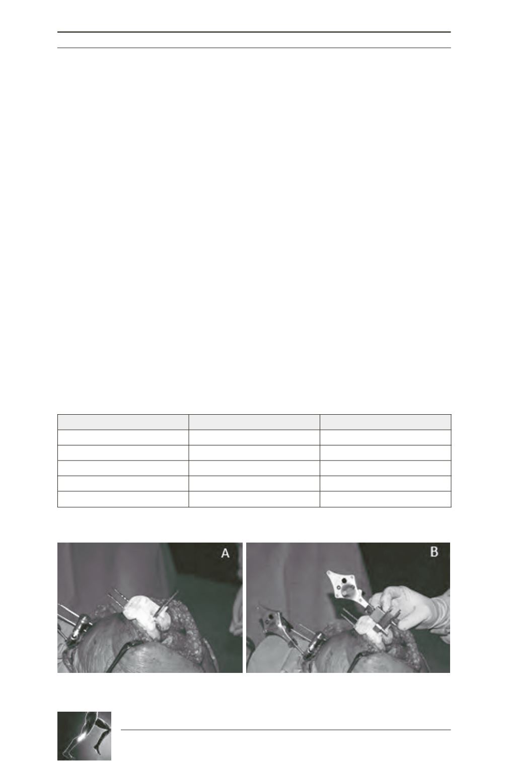
The PSCB was used in accordance with the
manufacturer’s instructions, with careful
positioning over the articular surfaces. The
accuracy with which the PSCB conformed to
the articular surface was checked by an observer
from Smith and Nephew, ensuring the PSCB
was used in the recommended fashion. Drill
holes and pins in the tibial and femoral
periarticular bone were placed through the
respective PSCB to determine the orientation
of the standard cutting guides, which were
recorded with the navigation system (fig. 1)
[2]. The parameters assessed intraoperatively
included the PSCB alignment and depth for
both the femoral (coronal, sagittal, rotational)
and tibial cuts. In addition, agreement between
the planned sizing for the tibial and femoral
components and the sizing determined
intraoperatively was recorded (Table 1).
The PSCB-defined bone cuts were only used if
the intra-operative measurements confirmed
acceptable alignment, which was defined as
within ±2° or ±1mm of the pre-operative plan
in each plane. If this was not achieved, the
PSCB was removed and the procedure was
performed with the navigation system in a
standard manner.
Fig. 1 : Intraoperative view of PSCB positioning for the femur (A)
and assessment of alignment with the navigation system (B).
General
Femoral PSCB
Tibial PSCB
Fit
Sizing
Sizing
Conformity
Coronal alignment
Coronal alignment
Limb alignment
Sagittal alignment
Sagittal alignment
Rotation
Cut depth
Cut depth
Table 1: Parameters assessed intraoperatively.


