
Evidence of Trochlear Dysplasia in Patellofemoral Arthroplasty Designs
59
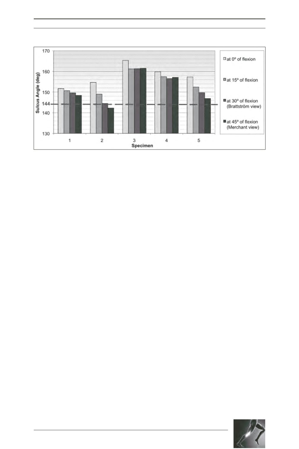
The height of the lateral facet of all trochlear
profiles is presented graphically (fig. 6). Visual
comparison reveals that the lateral trochlear
facet height is inversely proportional to the
sulcus angle. Three specimens had a facet less
than 5mm high through the entire range of
early flexion (0° to 30°), and two specimens
had a facet less than 5mm high beyond early
flexion (30° to 45°).
When projected onto the frontal place, the
trochlear groove was oriented laterally in all
specimens within the range 1.6º to 13.5º
(Table 1). The ratio of ML width to SI height at
different flexion angles reveals that in most
specimens the ML width is slightly greater at
15º and 30º of flexion than at 0º and 45º of
flexion. Two specimens were relatively narrow
(ML/SI < 1) while the other three specimens
were relatively wide (ML/SI > 1).
Discussion
This study revealed that contemporary PFA
implants are not always designed with anatomic
trochlear parameters, and some designs meet
the radiologic criteria of trochlear dysplasia.
Such components suppress intrinsic anatomic
features that are essential for normal patello
femoral tracking. Failures of PFA implants are
associated with two types of post-operative
complications: (
i
) late complications due to the
spread of arthritis to the tibiofemoral joint [36,
30, 13, 28, 24, 26], and (
ii
) early complications
due to patellar mal-tracking, including painful
instability, subluxation or dislocation [30, 13,
28, 24, 26]. Several studies reported improved
clinical outcomes for recent PFA models [13,
27, 28, 30, 35, 36], and many authors attributed
the reduced complication rates to enhanced
trochlear component designs, together with
better surgical techniques and instrumentation,
that enabled restoration of normal patellar
tracking [27, 28, 30, 35, 36]. The recent meta-
analysis by Dy
et al.
[13] affirmed that
complications of recent PFA models (14%) are
fewer than those of earlier PFA models (39%)
but still higher than those of TKA implants
(7%), and that most PFA complications remain
due to patellar instability and maltracking.
The surface geometry of the trochlear
component is of great importance, in addition
to accurate limb alignment and soft-tissue
balancing, to restore patellar kinematics and to
prevent patellar dislocation [28, 29]. In a
normal knee, the patella is guided into the
trochlear groove by the medial patellofemoral
ligament in early flexion [30, 31], and by the
lateral trochlear facet in later flexion [32, 33,
30]. In a knee with patellofemoral arthritis, the
Fig. 6: Height of lateral trochlear facet for all specimens at different flexion angles. The dashed red line
represents the radiographic indicator of trochlear dysplasia (lateral facet lower than 5mm in the “Brattström
View”).


