
M. Bonnin
72
How imaging the soft-
tissues around a TKA?
An In Vitro study
A precise imaging of the soft tissues tracking
around TKAs’ during knee flexion would be
very valuable but it is challenging due to their
Chromium-Cobalt alloy structure. Magnetic
Resonance Imaging (MRI) does not provides
good quality images in TKA, CT-scan analyzes
only components orientation, bone quality,
bone losses and components loosening.
Ultrasonography can also be used with TKA
for clinical purposes but it is hardly used for
precise anatomic investigations. Arthroscopy
can also be used but only in selected
indications.
The purpose of this study was to analyse
In
Vitro
the relationships between soft tissues and
TKA with commercially available implants.
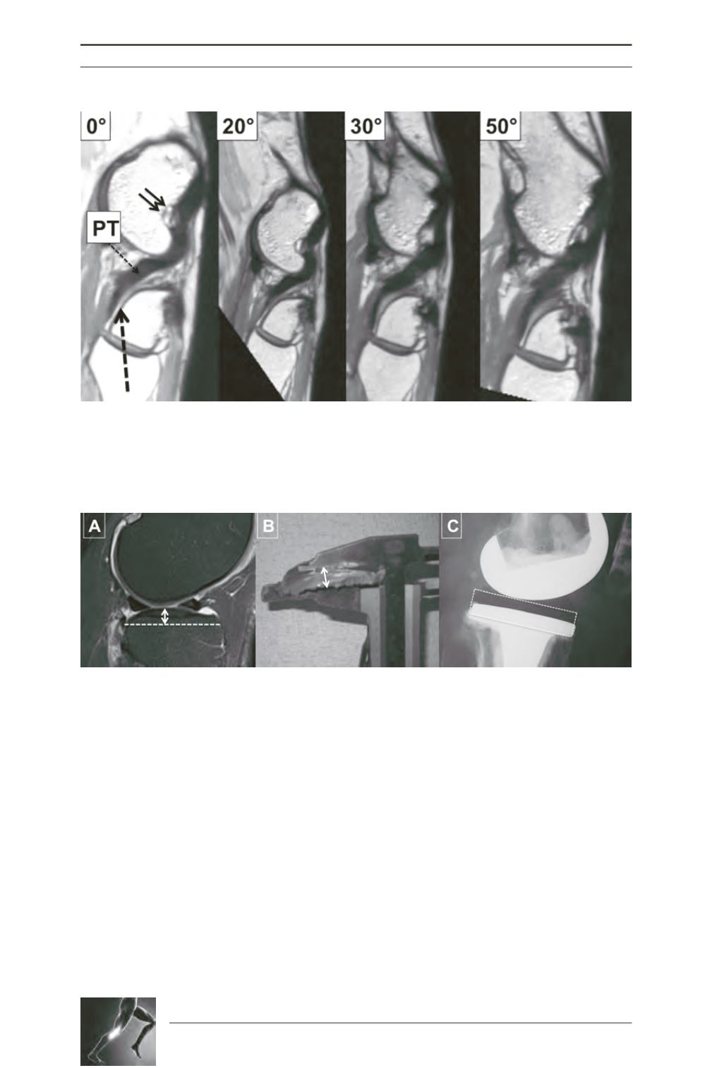
Fig. 1: These images were obtained with a 7 Tesla MRI from 0° to 50° of flexion. The slices are oblique and
aligned with the Popliteus tendon (PT) from its insertion at the level of the lateral epicondyle (double black
arrow) down to its contact with the lateral tibial plateau. In the posterolateral area, the PT is in close
contact with the lateral plateau through a cartilaginous surface (dotted black arrow).
Fig. 2: In the sagittal plane, the shape of the normal lateral plateau is convex (A from MRI). During TKA, the
level of resection is generally referenced to the lateral plateau, with a measurement at the top of the
convexity (B). The prosthetic tibial plateau is more or less a parallelepiped in the sagittal plane (C) and
therefore the posterolateral area has an increased volume compared with the native tibia.


