
Soft tissues and TKA
75
Consequently, if the contour of the tibial
component matches exactly the bony contour
of the tibial cut – theoretical optimal sizing –
the PT impinges on the polyethylene on a
significant area. Ideally the tibial plateau should
be undersized by 5 to 6mm in the posterolateral
corner of the tibia but this is difficult to satisfy
this goal with symetrical implants and
frequently the surgeon must accept a sizing
compromise (undersizing the medial tibial
plateau) or a position compromise (internal
rotation of the tibial baseplate).
This study demonstrate that optimal tibial
component design should be adapted to the
dimensions of this “functional” tibial plateau
(bone cut area without the PT contact area)
rather than to the raw bony contours. New
morphometric investigations should therefore
be conducted in order to redefine these
dimensions.
Imaging of the soft
tissues after TKA: The
modified tracking of
the PT
After TKA implantation the tracking of the PT
is greatly modified. This is true in case of
oversized plateau in the AP dimension but also
in apparently normosized implant (fig. 6). A
normal tracking of the PT was observed only in
specimens where the tibial component was
significantly undersized on the lateral plateau.
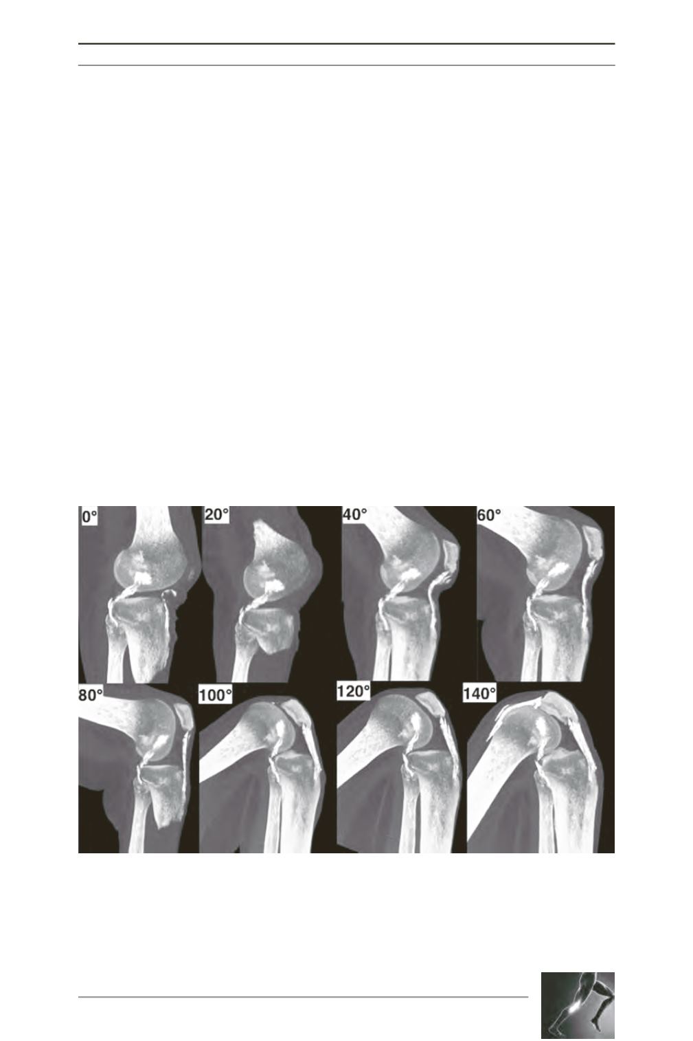
Fig. 5: These images are obtained from sagittal views of the normal knee during flexion, with thick slices
visualizing the all PT (OsiriX software). The PT is white, due to the baryum injection.


