
M. Bonnin
74
TKA (the contours of the implants is always
more than 2mm inside the bony contour) and
3)
Oversized TKA (the TKA overhangs more than
2mm from the bony contours).
Analysis of DICOM images
DICOM images were analyzed with OsiriX®
software, with 3D multiplanar reconstructions.
From these raw images segmentation was done
withMimics®software (Materialize®, Leuven,
Belgium) in order to obtain three-dimensional
images. To improve the quality of the implants
visualization, the Stereo Litography files (STL)
of the implants were obtained from the
manufacturer, so that we can match with the
raw DICOM images (fig. 3).
Imaging of the PT in the
normal knee: The
“functional” tibial
plateau and the
“anatomic” tibial
plateau
In a normal knee, the PT crosses the postero-
superior surface of the tibial plateau, with a
maximum overlapping of 5.5mm (75mm
2
),
observed while the knee is fully extended
(fig. 4 and 5). This overlap decreases from 0° to
90° of flexion. In deep flexion, the PT remains
distant from tibial plateau.
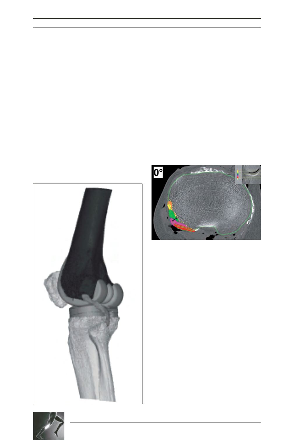
Fig. 3: Three-dimensional reconstruction of the knee,
obtained from DICOM images after implantation of
the TKA. In this specimen, the prosthesis was
“normosized”. The PT crosses the posterolateral
corner of the plateaus. Reconstructions were made
with Mimics® software (Materialize®) with a fusion
of the STL files of the implant.
Fig. 4: This axial slice represents the standard tibial
cut made at 9mm from the top of the lateral
plateau. The contours of the tibia are circled in
green. The positions of the popliteus tendon on
each CT slice, from 9mm (circled in green) to 5mm
above the joint line level (JL) (orange) are projected
on this slice (i.e., 7.5mm distal to JL in red, 5mm
distal to JL in purple, 2.5mm in blue, JL level in
green and 2.5mm above JL in yellow). The PT
overlaps the reference cut in all the posterolateral
area. The maximum overlap zone in this specimen
was 7mm (black arrow). To avoid impingement, the
prothetic plateau should no overhang from the
yellow dotted line, which mark the limit of the
“functional” tibial plateau.


