
41
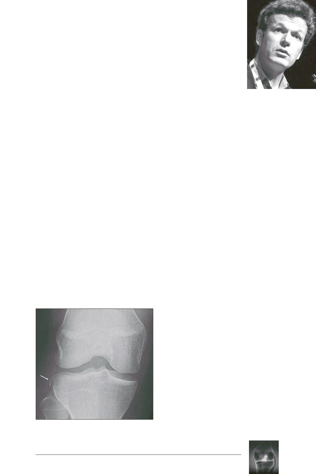
We now know that the Segond fracture is an
injury of the anterolateral ligament (ALL).
This small image of an avulsion involving the
lateral aspect of the tibial plateau is visible on
the AP view of the knee X-ray.
Pathognomonic for an anterior cruciate
ligament (ACL) tear, it is therefore an avulsion
of the ALL’s distal enthesis (fig. 1).
Seven teams have studied MR imaging of the
ALL since 2014 [1].
The ALL is certainly visible with MRI but the
diagnostic capabilities vary depending on the
different portions identified.
Some of these studies use 2D acquisition
protocols with a 3 to 4 mm section thickness. It
appears to be difficult to study a thin structure
of under 2 mm with sections of this thickness
and it is therefore logical to assume that certain
poor results are due to the acquisition protocols.
The ligament appears as a physiological
hyposignal. It is relatively relaxed on the MRI,
which is performed with the knee bent at 20° to
30° with a distal tibial enthesis curved on
coronal sections.
It is also possible to see the ALL’s meniscal
attachments, whose morphology varies.
For greater precision and efficacy, ideally 3D
1mm-section sequences should be taken. They
are available on all new 3 Tesla MRIs and on
the latest generation 1.5 Tesla MRIs. With
these sequences, the MPR mode can be used to
study the ALL much more precisely.
In routine practice, we use T2-weighted 3D
sequences without a fat suppression technique.
The ligament is clearly visible as a physiological
hyposignal relative to its surroundings. The
lateral inferior genicular artery is clearly
ANTEROLATERAL
LIGAMENT IMAGING
B. BORDET, J. BORNE, A. PONSOT,
P.F. CHAILLOT, O. FANTINO
Fig. 1:
AP view of knee X-ray. Segond fracture
(arrow)
.


