
B. BORDET, J. BORNE, A. PONSOT, P.F. CHAILLOT, O. FANTINO
44
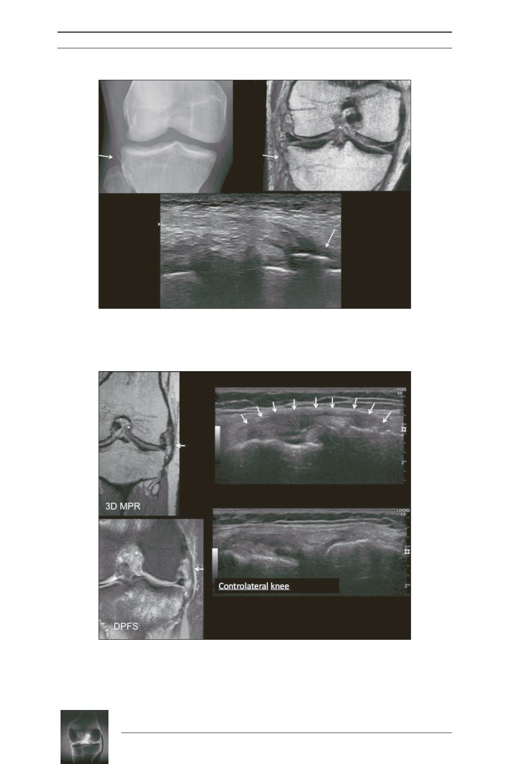
Fig. 5:
Example of ALL tear by distal avulsion
(arrow)
. The standard radiography
shows a Segond fracture. The 3D T2-weighted MRI and ultrasound show the
pathological thickening of the ALL and its bony avulsion at the distal enthesis. Note
the anterior cruciate ligament tear on the MRI
(asterisk)
. Thanks to Marie Faruch.
Fig. 6:
Example of an ALL tear without any bone injury. Severe damage to the ALL
under MRI and ultrasound without any bone injury. The ligament is completely
infiltrated and thickened under MRI with a clearly visible pathological hypersignal on
the sections using a fat suppression technique. Associated anterior cruciate
ligament tear
(asterisk)
. Comparative ultrasound sections clearly show the hypoechoic
ligament, which is thickened and distended on the injured side
(arrows)
.


