
ANTEROLATERAL LIGAMENT IMAGING
43
Thus, it is possible to look for a stretched and/
or torn ALL using ultrasound.
When an ALL injury is identified using MRI,
today we routinely conduct further explorations
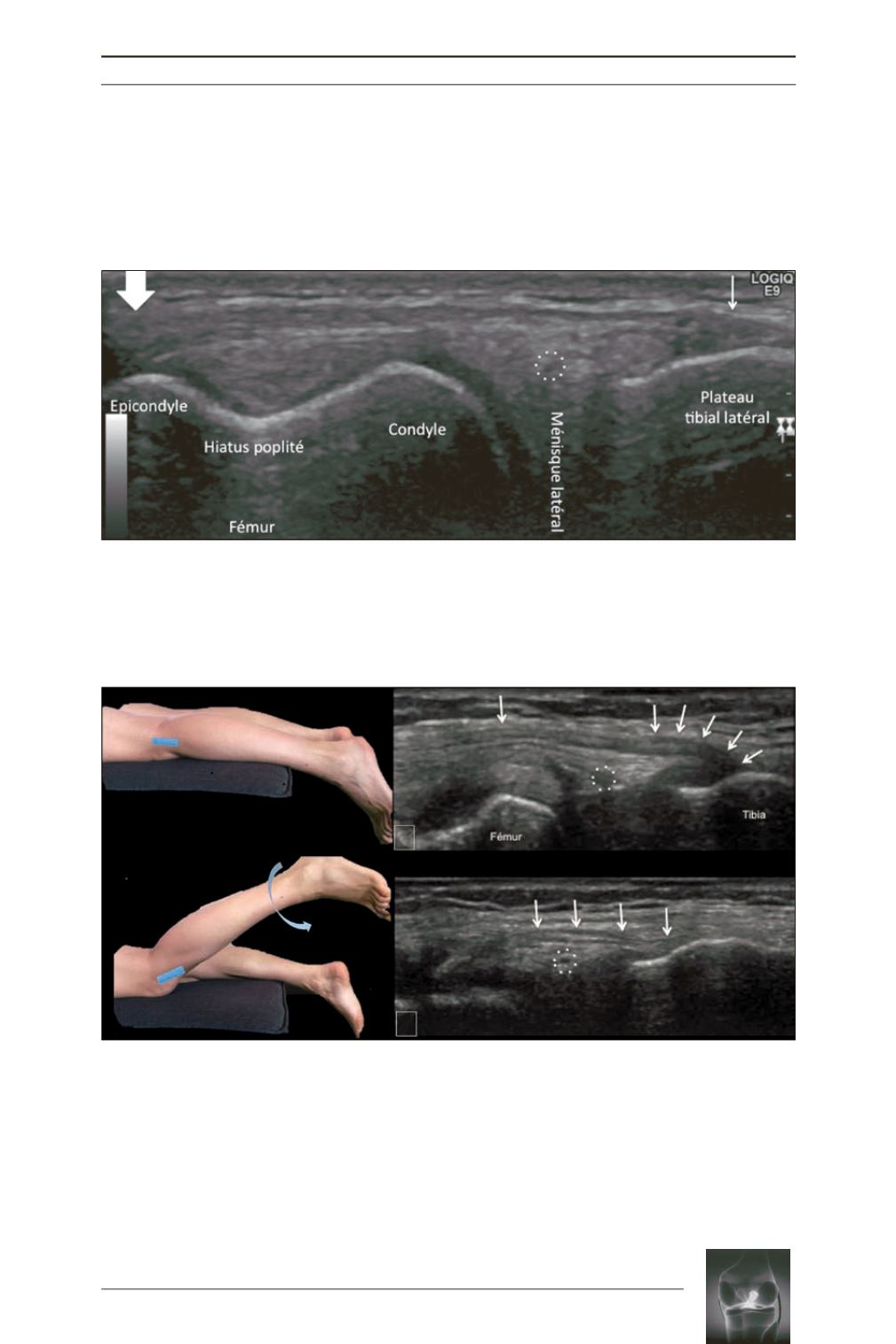
using dynamic ultrasound (fig. 5, 6).
Fig. 3:
Ultrasound of the ALL. Study with a high frequency 12 MHz superficial transducer. Normal
appearance in resting position in a longitudinal section. The ligament is thicker at its femoral enthesis
(large arrow)
and thinner at its tibial enthesis
(small arrow)
. It crosses the lateral inferior genicular artery
(dotted circle)
.
Fig. 4:
Ultrasound of the ALL. Study of the ligament (arrows) with a very high frequency 15 MHz superficial
transducer. Ligament in resting position
(figure a)
, relaxed at its tibial enthesis. Dynamic flexion and internal
rotation maneuver
(figure b)
making the ligament tense so that it straightens.
a
b


