
B. SONNERY-COTTET, R. ZAYNI
48
femoral and meniscotibial segments.According
to Hughston, this “capsular ligament” was
“strong and supported superficially by the
iliotibial band” and it played a significant role
in the knee’s anterolateral stability. In 1986,
Terry
et al.
[15] also referred to an anatomical
structure deep to the fascia lata that acts as an
“anterolateral ligament of the knee”. The
presence of this structure was confirmed by
Vieira [16] and then described in depth by
various teams [1, 7, 8].
ANATOMY
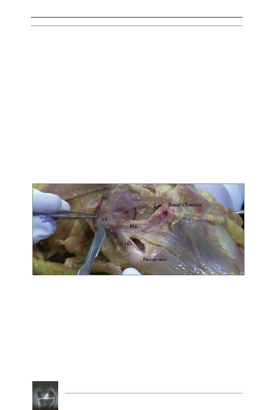
In 2016, we described a simple, reproducible
method to dissect and identify the ALL
surgically [4]. Dissection starts at the ALL’s
tibial insertion. Distal detachment of the biceps
femoral exposes the lateral collateral ligament
and also reveals the more superficial ALL.
Flexing the knee and maximally rotating the
tibia internally places tension on the ALL,
making it easy to identify. The ALL’s femoral
insertion has been the most controversial. The
current consensus is that it is located proximal
and posterior to the epicondyle [5, 6, 17, 18].
Near the joint line, the ALL has projections
on the lateral meniscus [8] and the
anterolateral capsule; most of its fibres fan
out and insert distally on the tibia between
the fibular head and Gerdy’s tubercle. Its
tibial insertion is more than 10mm wide [6].
It is located on average 21.6mm posterior to
Gerdy’s tubercle and 23.2mm anterior to the
fibular head [1], and is 10mm distal to the
joint line [1, 7, 8, 17].


