


 23 / 324

14
es
JOURNÉES LYONNAISES DE CHIRURGIE DU GENOU
22
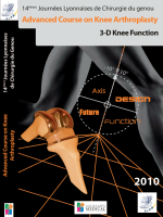
Fig. 3-A, 3-B: (Figs. reprinted, with permission, from: Marti RK, Kerkhoffs GM. Osteotomies
for malunions of the tibial head. In: Marti RK, van Heerwaarden RJ, editors. Osteotomies for
posttraumatic deformities. New York: Thieme; 2008. p 479-94.) Fig. 3-A Intra-articular cor-
rection is performed through the extended arthrotomy and the opening wedge osteotomy.
The depressed cartilage zone is marked circumferentially with a 2-mm drill-bit, either from
proximal as shown here or from distal through the proximal tibial osteotomy. Fig. 3-B After
the depressed plateau zone is marked with passes of a small drill-bit, the passes are connec-
ted with an osteotome before the complete zone can be elevated.
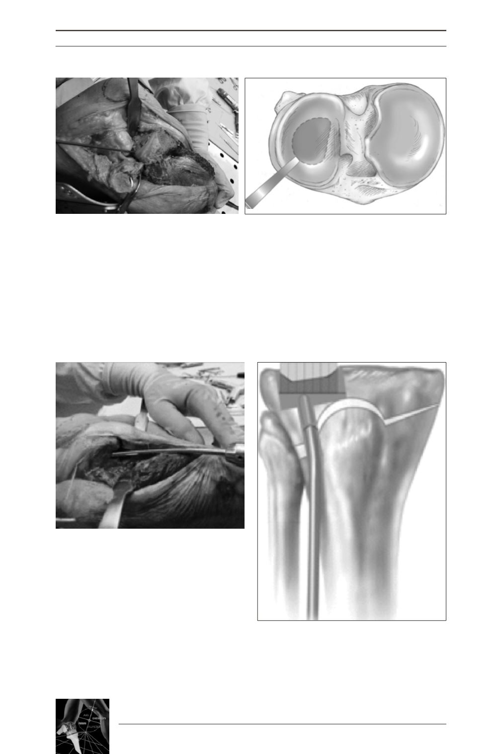
Fig. 4A, 4B :
4a: (Figs. Reprinted from: Kerkhoffs GMMJ et al. J Bone Joint Surg Am. 2009; 91 Suppl: 101-
1). With use of a curved impactor inserted through the window, the depressed area of the pla-
teau is elevated to conform to the lateral femoral condyle. 4b: To conform to the lateral femo-
ral condyle in both extension and flexion, an overcorrection of 1 mm is created. The correc-
tion is maintained by impacting cancellous autograft bone beneath the elevated segment
A
B
A
B