
E.A. Arendt
122
establish a short docking station for the MPFL
by over-drilling the K-wire with a 4mm
cannulated reamer for a length of 10mm. Even
if one’s preferred surgical technique involves a
different kind of patella fixation, this technique
can be used by inserting a K-wire as described
above and later removing it.
The author’s preference is to lengthen, not
release, the lateral sided soft tissue structures
of the patella (fig. 4). In support of lateral sided
lengthening: it is a more precise balance of the
patellofemoral forces and it reduces the
potential for excessive medial patellofemoral
translation. Conversely, lateral retinacular
lengthening does require either a larger incision
or two incisions. It slightly increases the
operating room time. At times, the gap between
the lateral structures is too great to be lengthened
and a release is necessary; however in the
surgical profile detailed above (“isolated”
MPFL reconstruction), the gap is rarely
>20mm. When one performs a lengthening of
the lateral tissue, one often has 15-22mm of
tissue to lengthen.
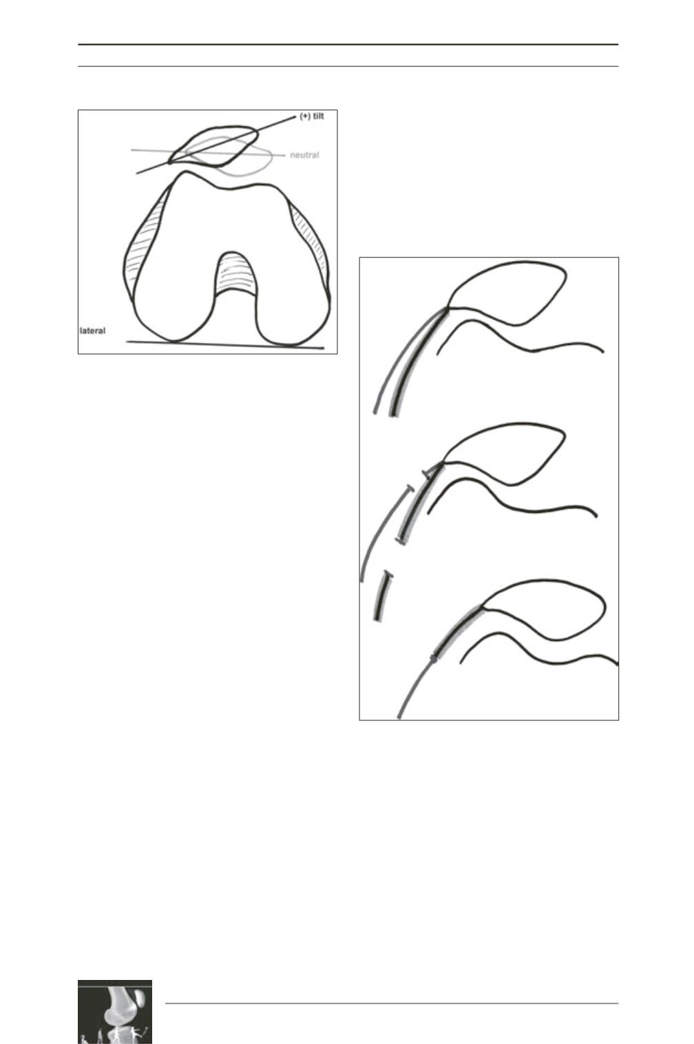
Fig. 3 : K-wire placed thru the M-L axis of the
patella intra operatively.
Black: patella M-L axis cannot be brought to a
horizontal level.
Grey: post-lateral retinacular lengthening/release.
Fig. 4 : Schematic drawing of lateral retinacular
lengthening : ITB = Iliotibial band, LPFL= lateral
patellofemoral ligament
Dark-grey: ITB
Light-grey: LPFL


