
Anatomical double bundle mpfl reconstruction
127
Preparing the femoral insertion site
To avoid non-physiological patellofemoral
forces, the femoral MPFL insertion has to be
very accurate. Therefore, a guide wire with an
eyelet is placed slightly posterior to the
midpoint of the medial epicondyle and the
adductor tubercle and the entering point into
the bone is marked with a clamp [10]. Then the
guide wire placement is controlled by a picture
intensifier on a straight lateral view to obtain
the correct anatomical femoral insertion; if the
graft is placed too anterior or proximal,
abnormal graft tensioning will lead to increased
patellofemoral pressures during flexion [3].
Therefore, we use the radiographic landmark
of the anatomical MPFL insertion which has
been shown to be located slightly anterior to an
elongation of the posterior femoral cortex in
between the proximal origin of the medial
condyle and the most posterior point of
Blumensaat’s line [6]. If necessary, the guide
wire entry point is corrected before overdrilling
to the contralateral cortex with a drill diameter
1mm larger than that of the graft loop.
Preparing the patellar insertion site
To achieve aperture fixation at the patellar side,
the free graft ends have to be fixated directly to
the patella. Therefore, the medial patellar
margin is prepared and two guide wires are
drilled tangentially into the patella at the
proximal and distal end of the medial edge. The
guide wires are subsequently overdrilled with a
cannulated 4mm drill to a depth of 20mm.
Graft fixation
The two free sutured graft ends are fixed into
the patellar holes one after each other, using a
4.75x15mm Swivel Lock (Fa. Arthrex),
achieving a direct anatomical graft fixation. To
accomplish this, the graft sutures are pulled
through the PEEK eyelet of the Swivel Lock,
and pushed into the drill holes. Keeping the
suture under tension, the graft ends are fixed
with the 4.75x15mm Swivel Lock screw
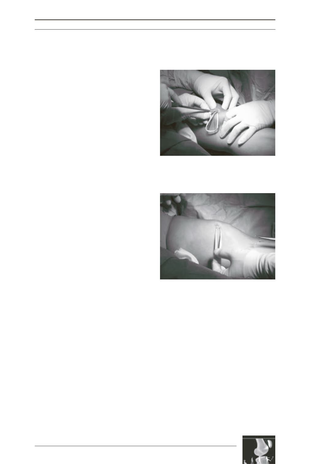
(fig. 2). In this way, a double bundle aperture
fixation at the patellar side is achieved, leaving
the graft loop free (fig. 3).
The suture loop is then used to pull the graft in
between layer 2 and 3 to the femoral insertion.
Next, a Nitinol wire is inserted into the femoral
drill hole and the suture loop of the graft is
pulled laterally using the guide wire. Finally,
while maintaining equal tension on both
bundles, the graft is pulled into the femoral
socket. Since biomechanical studies have
shown that the MPFL has its maximal length
and restraint against patella lateralisation in
30° of flexion (fig. 4) [3], femoral fixation is
performed in 30° of flexion with the lateral
patellar edge positioned in line with the lateral
trochlear border using a bioresorbable
interference screw. An anatomical femoral
insertion avoids an overcorrection, since an
Fig. 2: Using two Swivellock anchors, the armed
graft ends are inserted and fixated into the
patella.
Fig. 3: Graft ends are fixed using a 4.75x15mm
Swivel Lock screw while tensioning the graft.


