
P.B. Schoettle
128
overtension of the graft can only occur if the
femoral tunnel is placed too far anterior or
proximal. In this case, the insertion point would
move towards posterior in flexion, leading to a
lengthening of the distance between patellar
and femoral insertion, increasing the load onto
the graft, and consequently, onto the patello
femoral joint.
If adequate medial restraint has been restored,
lateral patellar dislocation should no longer be
possible and routine skin closure is performed
after reattaching the aponeurosis of the VMO
back to the medial edge of the patella with
resorbable sutures.
Postoperative
treatment
Compared to other techniques, this aperture
fixation with a biotenodesis screw at the patellar
insertion provides an immediate stable tendon
to bone fixation with an ultimate load to failure
force at the patellar side higher than the 208N
needed to rupture an intact MPFL [3]. Weight
bearing is allowed, however, no more than
20kg until wound healing, while leg raising
and quadriceps setting exercises can be started
immediately with a free range of motion as
tolerated.
Low impact activities such as running or
cycling are allowed at 6 weeks post-op; full
activity is permitted at 3 months.
Discussion
The most important finding and improvement
in using the above described technique was the
possibility of an immediate full range of motion
due to the aperture fixation at both sides. The
benefit of anatomic graft positioning in ligament
reconstruction has been known for a long time
and has been clearly demonstrated in ACL
reconstruction. Anatomical reconstruction of
the MPFL is particularly important as
biomechanical studies have demonstrated that
the length change pattern of a MPFL
reconstruction depends critically on the site of
the femoral attachment; moreover kinematics
change significantly when the patellar or the
femoral insertion has been off by only 5mm
[4]. Aside from tunnel placement, graft fixation
is the other determining factor in ligament
reconstruction [7]. Non-aperture fixation at
either the femoral or patellar insertion can
increase the risk of a delayed or insufficient
tendon to bone healing, which may result in
early loosening or slackening of the graft. To
avoid this, a restricted range of motion is
recommended by some surgeons; however, this
may lead to arthrofibrosis, potentially
necessitating an additional arthroscopic
arthrolysis, carrying the additional risk of
damage to the graft.
However, until today, only one technique
describes a double bundle aperture fixation at
both sides where the graft is looped through a
bone tunnel in the patella [9]. In this technique,
the graft is shuttled through the patella and
fixated press fit without any fixation devices,
providing a high intial fixation strength.
The aim of this manuscript was therefore to
describe a procedure for an anatomical double
bundle reconstruction of the MPFL, respecting
not only the ligament shape and both the
anatomical patellar and femoral insertion areas,
but also an aperture fixation.
In recent studies, a tendon transfer is described
either to the patella or to the femur for
reconstructing the MPFL [11, 12]. However, in
these techniques, not only the is transferred
muscle weakened in its original motion, but
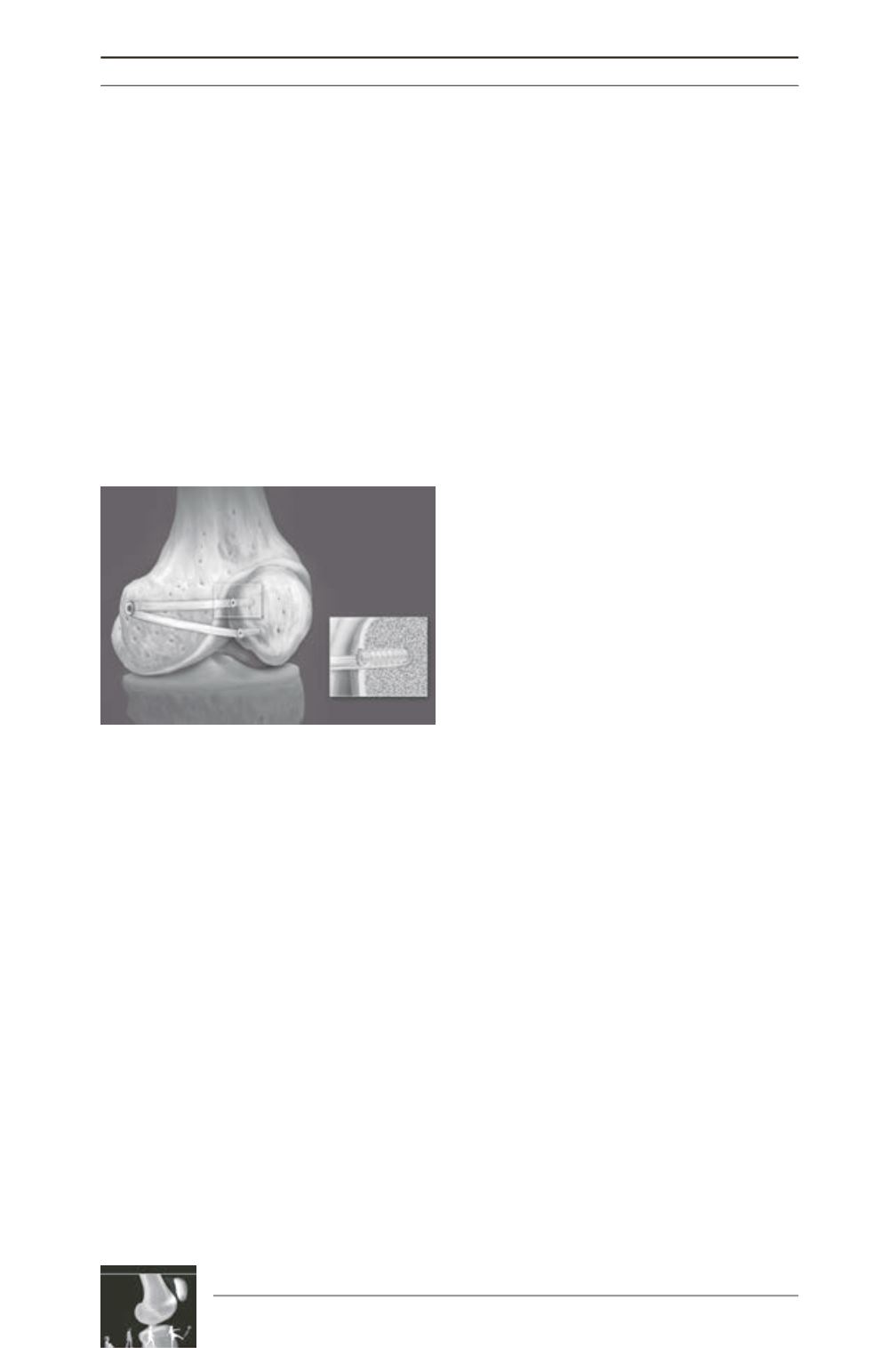
Fig. 4: The double bundle aperture fixation is
achieved at the patellar side as well as at the
femoral insertion.


