
All Surgical Procedures (video) - Patellar tendon procedure
137
Postoperative
treatment
Rehabilitation is started at the time of the first
outpatient return visit. The individual keeps the
brace on for three weeks, during which
isometric exercises are started and analgesia
(cryotherapy) and electrostimulation are
administered. We initially advise patients to
use a pair of crutches and walk without
loadbearing on the knee, and to perform
cryotherapy at home. During the first week, the
load will gradually be applied, up to the
patient’s pain limit. Starting in the first to third
week, we add in larger gains in range of motion,
after the third week, the immobilizer is keep off
and bicycle are use with loading and initial
proprioception exercises. From the sixth week
onwards, we start closed kinetic chain exercises
and gradually start to open kinetic chain
exercises. Our aim is that patients should be
able to return to their preoperative sports
activities after approximately 10 to 12 weeks.
Future possibilities for
the technique
We are seeking to adapt the skin incision, so
that the result is more cosmetic.
Abstract
In 2001 we presented a proposal for a surgical
technique to reconstruct the Medial Patello
femoral Ligament – MPFL using the patellar
tendon and in 2007 we published the technique
[1].
The technique consists of dissection to reach
the peritendon of the patellar ligament, taking a
graft from the medial third of the patellar
tendon to reconstruct the Medial Patellofemoral
Ligament. By means of a subperiosteal incision,
we release the distal extremity of the tendon
from the anterior tuberosity of the tibia and flip
this strip, medially and superiorly. Initially, we
perform a subperiosteal release on the patella,
as far as the junction of proximal third, with the
medial third as a graft positioned at femoral
insertion site and fixed using an interference
screw within femoral tunnel. If the graft is too
short, there is a possibility of not making the
tunnel and fixing it with absorbable or non-
absorbable anchors.
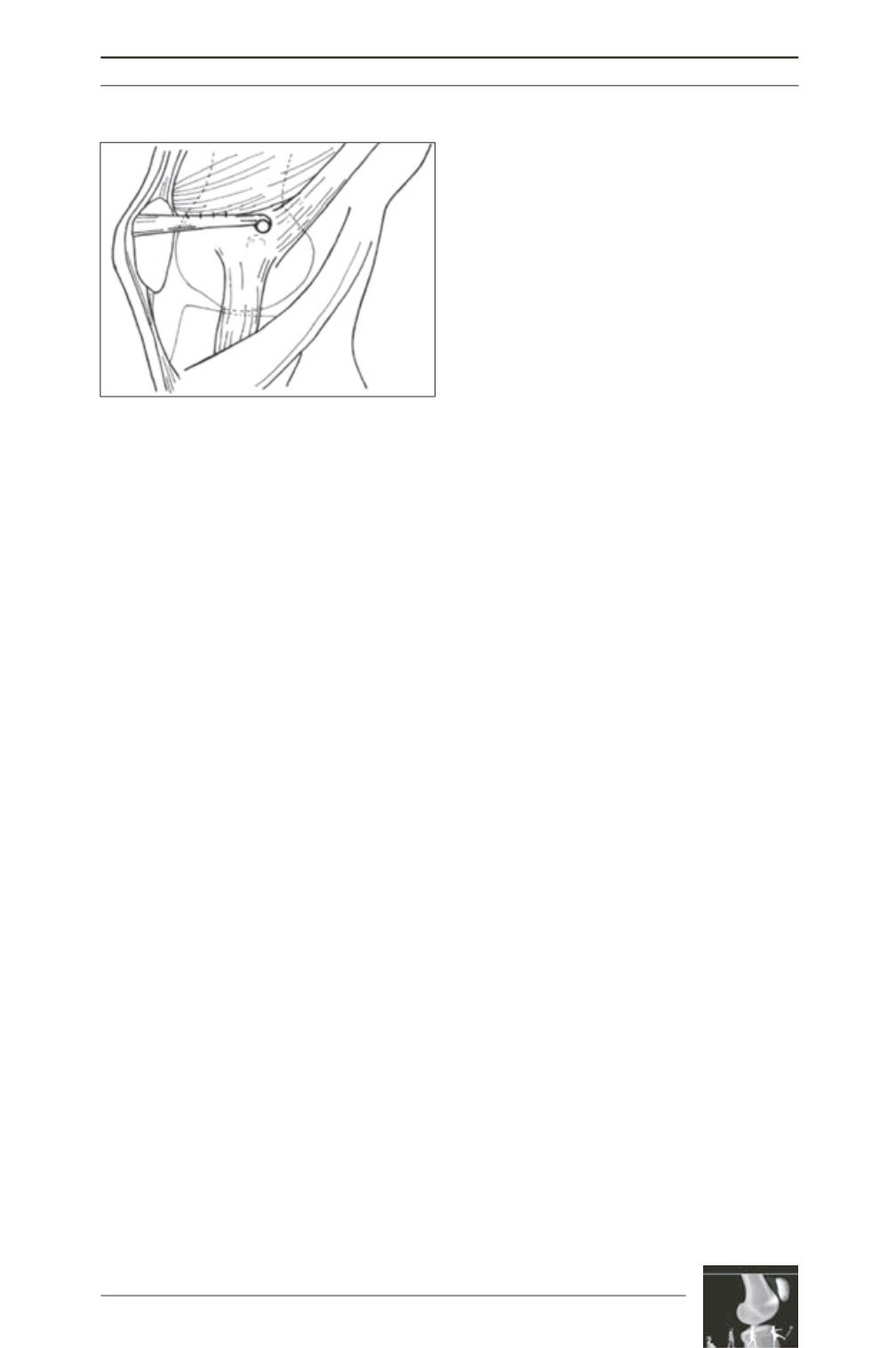
Fig. 4 : Suture of the vastus medialis muscle to the
ligament graft
Literature
[1] Camanho GL, BitarAC, HernandezAJ, Olivi
R. Medial patellofemoral ligament reconstruction: a novel
technique using the patellar ligament.
Arthroscopy 2007,
23(1): 108.e1-4.


