
G.L. Camanho
136
We perform layer-by-layer dissection to reach
the peritendon of the patellar ligament, as done
when taking a graft from the patellar tendon to
reconstruct the anterior cruciate ligament. The
peritendon will be incised vertically but
medially, since only the medial portion of its
structure will be used. We use the medial third
of this tendon.
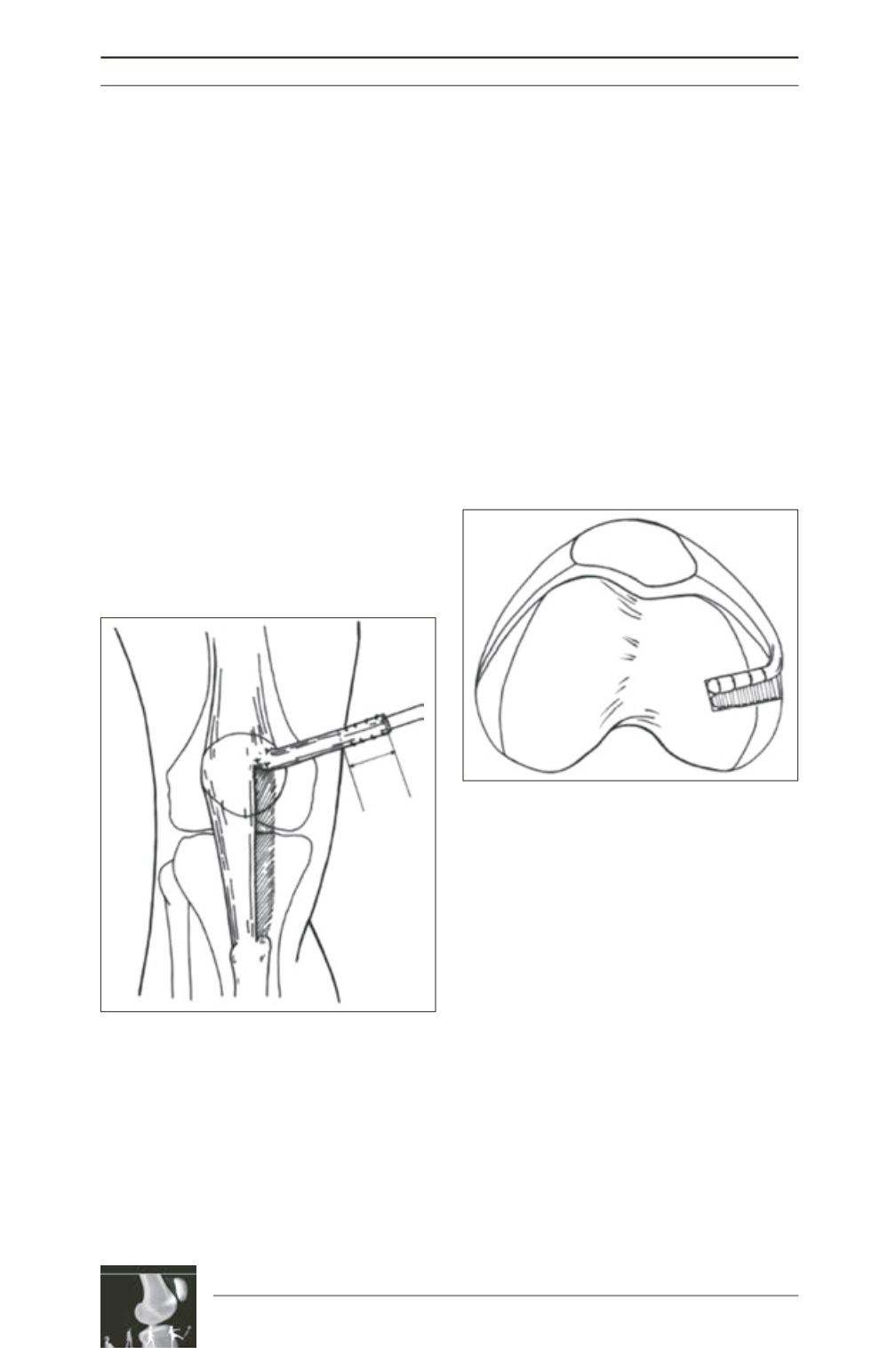
By means of a subperiosteal incision, we
release the distal extremity of the tendon from
the ATT and flip this strip, medially and
superiorly. Before this, however, with the
purpose to reproducing the anatomy of the
MPFL at its patellar insertion, we perform
subperiosteal release on the patella, as far as
the junction of the proximal third with the
medial third (fig. 2). We then perform
reinforcement using resistant thread, suturing
the graft to the extensor apparatus, which is
inserted into the patella.
The femoral insertion covers a more diffuse
area and is posterior and proximal to the medial
epicondyle. At this point, we need to measure
the distance to the patellar insertion and the
graft fixation point on the femur. We perfom
Krackow suturing on the free end of the tendon
and position the graft at the femoral insertion
site, allowing the entrance of the stitches into
femoral tunnel to further fixation with
interference screws. Next, we make the tunnel
using a drill bit of the same size as the graft is
ad at the end using an absorbable interference
screw (fig. 3). If the graft is too short, we have
the possibility of not making the tunnel and
fixing using absorbable or non-absorbable
anchors. The fixation should be done at
60 degrees of flexion, the position at which we
apply tension to the ligament.
It is of prime importance at this point to be
concerned about testing the total flexion and
stability of the fixation method.
We complement this technique by developing a
dynamic system between the reconstructed
ligament and the lower border of the vastus
medialis muscle, by means of stitches to suture
the vastus medialis to the MPFL (fig. 4).
After washing the wound, we close the
peritendon using V
icryl
® 3.0 and then close
the wound using intradermal stitches of
colorless M
onocryl
®. We do not routinely use
a drain and we prefer removal using the
tourniquet and perform hemostasis.
We perform infiltration using local anesthetics
in the skin incisions and we finish by bandaging
and immobilizing using an inguinal-malleolar
brace.
Fig. 2 : Diagram showing removal of the medial
third of the patellar tendon
Fig. 3 : Diagram showing fixation in the femoral
insertion using interference screw


