
C. Lapra, S. Chomel, M. Bakir
226
Many studies do not show any clear evolutive
link
between
the
cartilage
disease
“chondromalacia patellae” and patellofemoral
arthrosis. However, in athletes, PF cartilage
lesions are considered as related more to trauma
or micro-trauma than to over-use [3]. Dupont
[12] considers that cartilage lesions in children
and athletic teens are more frequently unnoticed
than they are actually rare [11].
As viscosupplementation has proven effective
in common arthrosis, authors suggest wider use
in athletes as soon as patellofemoral chondral
lesions appear in athletes; hence the usefulness
of detecting such lesions early.
What can be expected from a
patellofemoral MRI focused on the
cartilage?
Due to its superficial location and the thickness
of its cartilage, the patellofemoral joint lends
itself well to MRI evaluation and it is the joint
whose cartilage has been studied most.
The purpose of the MRI sequences we have
is to:
- clearly demarcate the joint surface of the
trochlear and patellar cartilage,
- define the thickness of the cartilage,
- explore the deep layer and the osseous
portion: tide-mark and subchondral bone,
- differentiate the cartilage from the intra-
articularfluidtodetectsuperficialirregularities
or notches of varying depths that may reach
the subchondral bone,
- determine the biochemical composition of the
lamellar structure of the cartilage so as to
detect intra-articular anomalies, without
surface lesions.
Contribution of 3D MRI sequences:
- What are the specifications for an MRI
sequence? The “ideal” MRI sequence would
be the one that combines short examination
time, a good signal-to-noise ratio, very good
spatial resolution, 3D study enabling ultra-
fine sections and multiplanar reconstructions,
specificsignalfromthecartilagedifferentiating
it from the articular fluid, specific signal from
structural modifications. Like any technique,
MRIs are a compromise!
- 3DFast Spin-Echo (FSE) sequences including
submillimeter sections have been improved
thanks to so-called parallel imaging enabling
shorter acquisition times and improved spatial
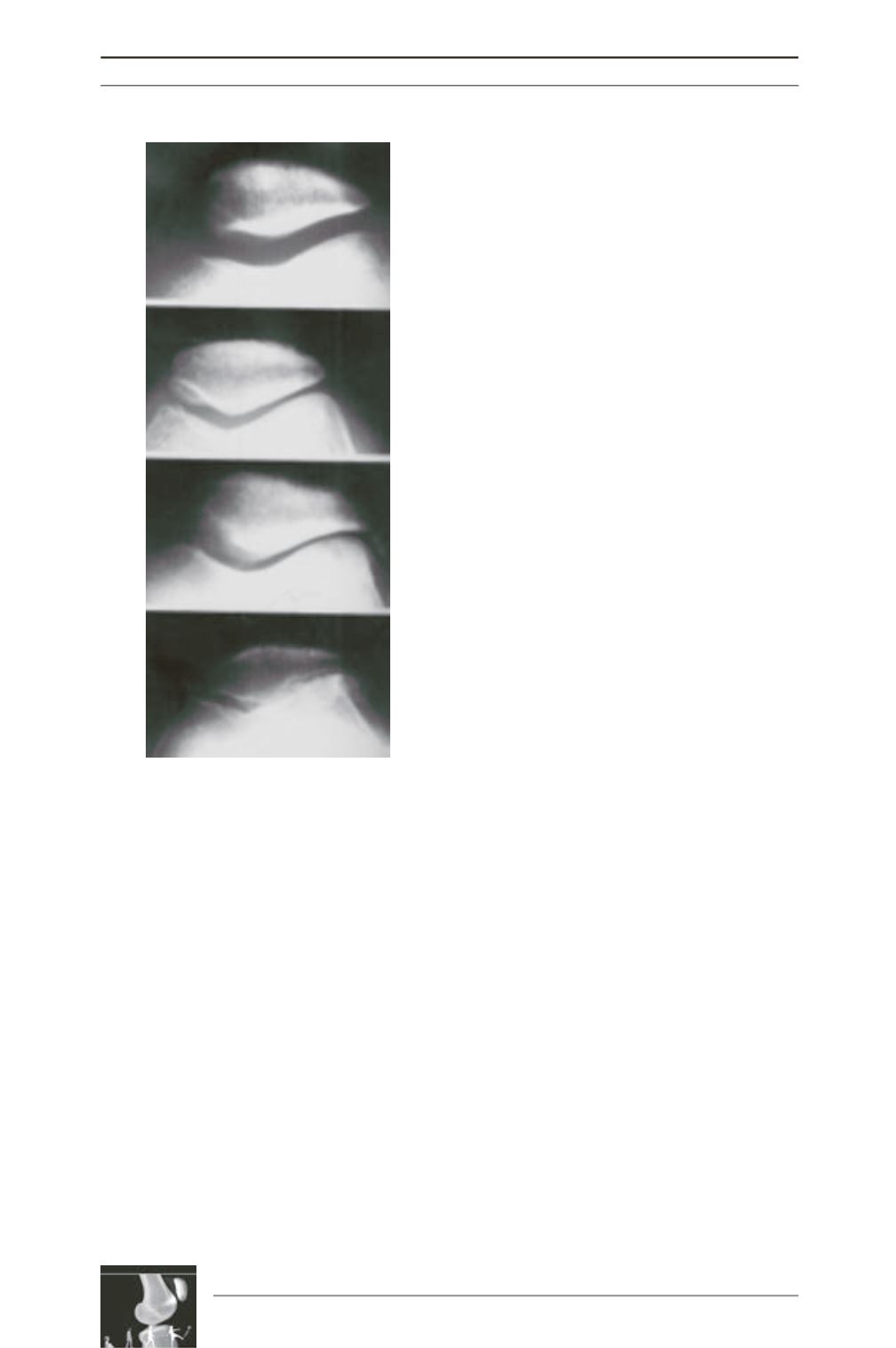
Fig. 1: Iwano classification
(source :
traité de chirurgie du genou, Ph. Neyret,
G. Demey, E. Servien, S. Lustig).


