
C. Lapra, S. Chomel, M. Bakir
232
- On CT scan or MRI, a deficient lateral
trochlear slope or insufficient trochlear
depth, the lateral trochlear inclination index
can be both measured on CTsan or MRI [4].
• Patella alta on the lateral X-ray,
• Lateral tilt of the patella (axial, lateral view,
MRI or CT-scan measurement of the patel
lar tilt),
• Measurement of the TT-GT distance on CT-
scan or MRI [15, 31] (fig. 14 a,b).
Trochlear dysplasia can be studied by CT-scan
or MRI [4, 17, 23, 31], different studies validate
the instability measurements validated with the
CT-scan or MRI. Thanks to its 3D reconstruc
tions, scans make it possible to thoroughly
explore the trochlear anatomy and the insufficient
depth of the trochlear groove.
To conclude
, imaging assessments must be
capable of answering two main questions:
- Inthecaseofpainindicatingthepatellofemoral
joint, are there degenerative chondral lesions,
or even proven osteoarthritis? If so, what is
causing the pain: a subchondral edema?
Lateral patellofemoral friction? Associated
synovitis?
- Are there anatomical predispositions to
instability? The traditional criteria shown on
the lateral view (trochlear dysplasia, patellar
tilt, patellar height) were also found on the latest
generation MRI and 3D and 2D CT scans.
The “best radiological exam” will undoubtedly
remain the most exhaustive in terms of
information provided for the therapeutic
treatment. As such, MRIs have become almost
mandatory!
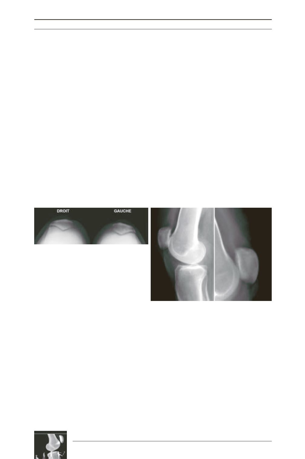
Fig. 14a: Patellofemoral Joint OA on axial view
Fig. 14b: trochlear dysplasia
Literature
[1] Arendt EA, Fithian DC, Cohen E. Current
concepts of lateral patella dislocation.
Clin Sports Med. 2002
Jul;21(3): 499-519. Review.
[2] Barbier-Brion B, Lerais JM,AubryS, Lepage
D, Vidal C, Delabrousse E, Runge M, Kastler
B. Magnetic resonance imaging in patellar lateral femoral
friction syndrome (PLFFS): prospective case-control study.
Diagn Interv Imaging. 2012 Mar; 93(3):e171-82.
[3] BrittbergM, Winalski CS. Evaluation of cartilage
injuries and repair.
J Bone Joint Surg Am. 2003; 85-A Suppl
2: 58-69.
[4] Carillon Y, Abidi H, Dejour D, Fantino O,
Moyen B, Tran-Minh VA. Patellar instability:
assessment on MR images by measuring the lateral trochlear
inclinaison. Initial experience.
Rgy 2000 Aug; 216(2): 582-5.
[5] Caton J. Method of measuring the height of the patella.
Acta Orthop Belg. 1989; 55(3): 385-6.
[6] Cotten A, Boutry N, Demondion X, Bera-
Louville A. Imagerie musculosquelettique : Pathologies
locorégionales.
Masson mars 2008.
[7] Chu CR, Williams A, Tolliver D, Kwoh CK,
Bruno S 3
rd
, Irrgang JJ. Clinical optical coherence
tomography of early articular cartilage degeneration in
patients with degenerative meniscal tears.
Arthritis Rheum.
2010 May; 62(5): 1412-20.


