
C. Lapra, S. Chomel, M. Bakir
230
Delayed gadolinium-enhancedMRI of cartilage
(dGEMRIC)
[30]: these sequences consist of
an intra-articular injection of a double dose of
gadolinium, associated with the patient walking
for 15 minutes. The examination is done after
90 minutes. It is a “molecular” study of the
cartilage that is not routinely done. In healthy
cartilage, gadolinium (Gd) does not penetrate
cartilage; Gd fixation increases if the
glycoaminoglycan content decreases.
Associated lesions: synovitis, subchondral
bone edema
Many studies have shown that the pain of
arthrosis was often linked to secondary lesions
associated with edema of the subchondral bone
due to trabecular bone microcracks and
synovial inflammation, which explains the
evolution in spurts [14, 18, 32].
Synovitis
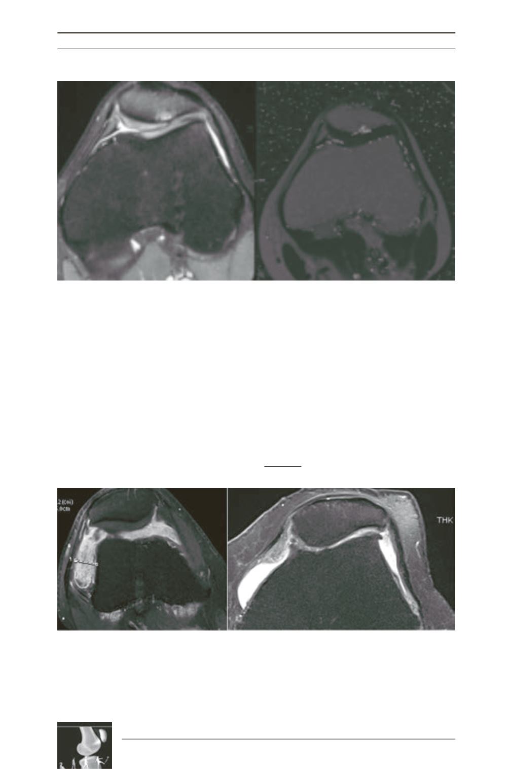
(fig. 11)
Fig. 10: There is cartilage thinning of the lateral patellar facet, not visible on the T2 map.
Fig. 11: Same patient, MRI. - a: Right knee MRI with injection: synovitis. b: On the left knee (Proton density),
the joint space is more impaired but there is less synovial inflammation.
a
b


