
Imaging of Patellofemoral Joint Osteoarthritis
231
Subchondral bone
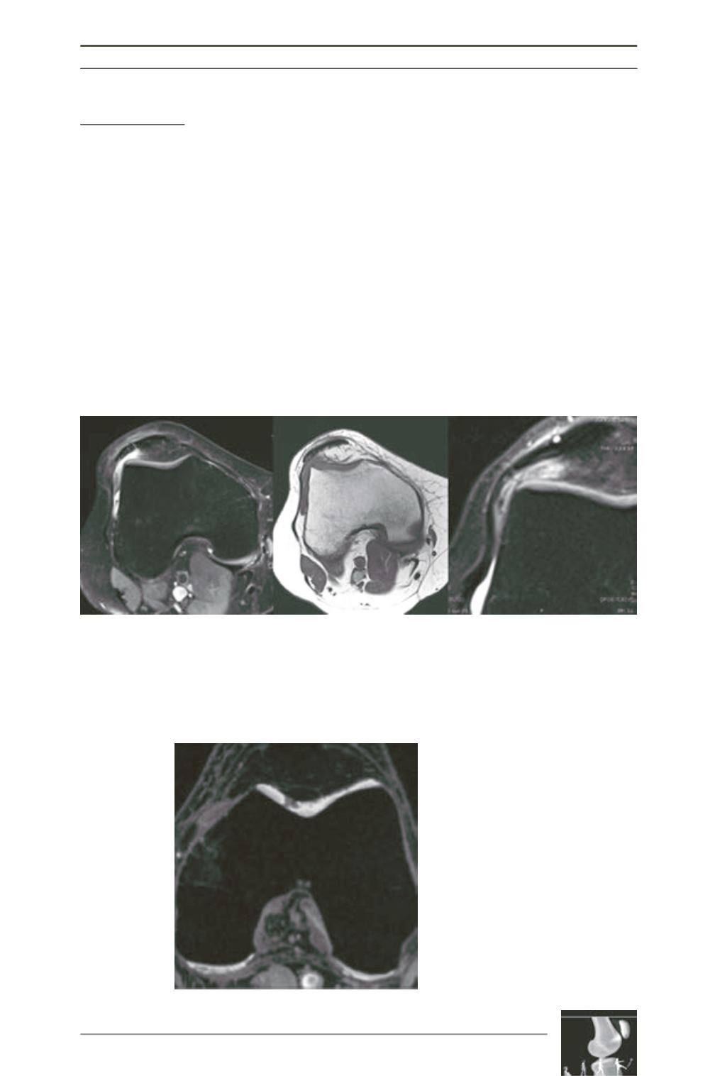
A lateral patellofemoral friction syndrome may
also be associated with degenerative cartilage
lesions, pre-disposed by a pre-existing morpho
logical anomaly of the joint, fostering
instability. This syndrome has also been
described as responsible for anterior knee
pain, in the absence of arthritic lesions [2, 8,
26] (fig. 12).
Future areas of development for cartilage
MRI will move towards ultra-high-field
(7 Tesla) MRIs, T1 mapping, molecular
imaging (sodium)
(fig. 13)
Question 2:
Is there an
anatomical predisposition
to instability?
Trochlear dysplasia is a frequent predisposing
factor for lateral patellofemoral; it is present
in 80% of the cases of patellofemoral OA.
Articular morphology findings are especially
important for the treatment of patellofemoral
OA.
The imaging assessment looks for:
• Signs of trochlear dysplasia:
- On the lateral view, crossing sign,
- Trochlear boss,
Fig. 12
Fig. 13: T1 Mapping
(source : Siemens)


