
Imaging of Patellofemoral Joint Osteoarthritis
227
resolution. The result of the sequences is to
acquire one plane and then, like with a scan,
create reconstructions in other planes. To
create high-quality multiplanar reconstruc
tions, MRI sequences must be as isotropic as
possible.
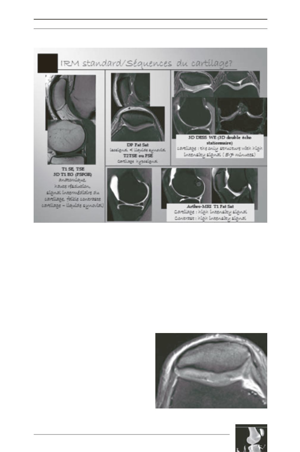
Comparison of the cartilage signal
based on sequences used [10, 24, 28]
(fig. 2)
As a result, several analyses can be
used to study the cartilage
- Morphological analysis, which can be
compared to Outerbridge [22] and Inter
national Cartilage Repair Society (ICRS)
arthroscopic classifications,
- Volume analysis,
- More recent “biochemical” analysis.
Morphological analysis
(fig. 3, 4, 5)
Exams with injections: arthro CT-scan (fig. 6a
and b).
MRI Arthrography (fig. 7 a,b): although the
intra-articular injection may sometimes make it
easier to grade the chondral anomalies, it makes
the exam more invasive.
Fig. 2: Source : Chomel S MD Imaging Cartilage of the knee, EPU January 2012 Lyon.
Fig. 3: ICRS Grade 2


