
Approach and Patella in Total Knee Arthroplasty
325
criteria for the lateral approach were the
presence of valgus knee or lateral subluxation
of the patella (whatever the frontal axis). The
prosthesis used was NexGen LPS Flex
(Zimmer) with a fixed, symmetrical tibial
baseplate. Rotation of the femoral part was
adapted by navigation to measurement of the
posterior condylar angle by pre-operative scan.
Rotation of the tibial baseplate sought to
position the tibial tray in parallel with the
femoral implant in complete extension (self-
adjustment). Rotation of the tibial implant was
not navigated.
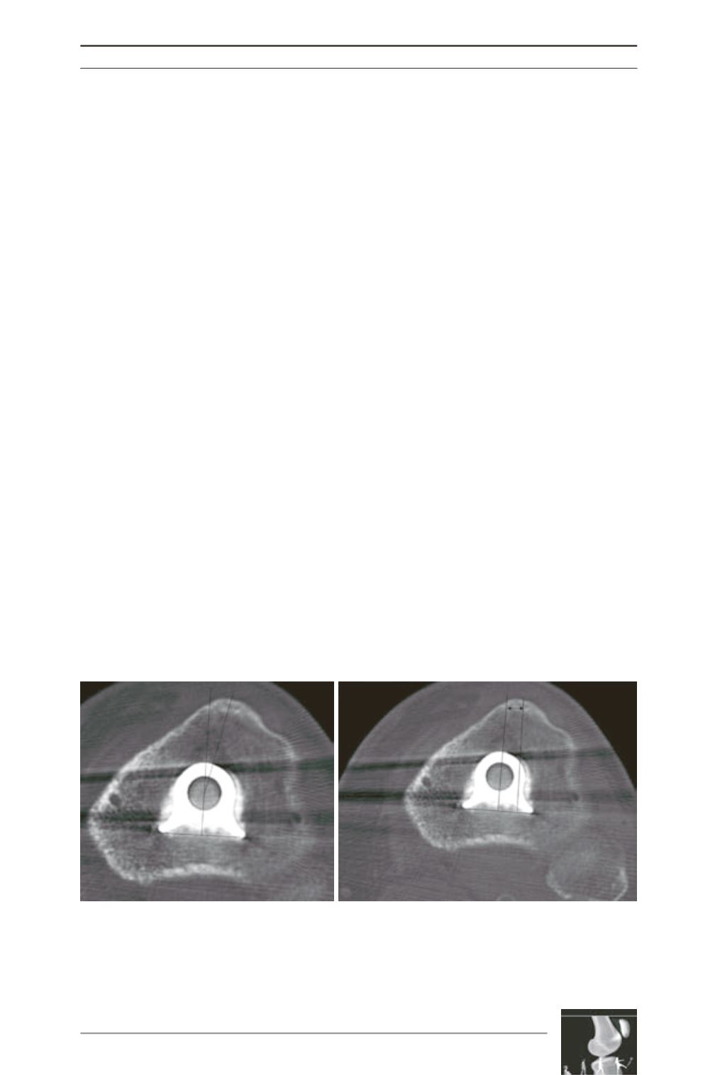
The post-op CT scan protocol for the tibia
aspect consisted of measuring (fig. 4):
On one hand, the angle formed by a line perpen
dicular to the transversal axis of the tibial
baseplate passing through its center and the line
linking this center to the middle of the ATT.
On the other hand, the distance between 2 lines
perpendicular to the transversal axis of the
baseplate, one passing through the center of the
baseplate and the other through the middle of
the ATT (SFHG protocol). Two measurements
could then be taken. In the medial group, the
average distance between the center of the ATT
and the center of rotation was +7.3mm
(minimum 1, maximum 16mm), and the
average angle of rotation was +18.8° (internal
tibial baseplate rotation) (minimum +1°,
maximum +36°).
In the lateral group, the average distance
between the center of the ATT and the center of
rotation was +1.4mm (minimum -5, maximum
+7mm) and the average angle of rotation was
+2.11° (internal rotation) (minimum -14°,
maximum +19°). In 6 out of 15 cases, the tibial
baseplate was in external hyperrotation: the
ATT was medial in comparison to the middle
of the baseplate. The lateral approach led to
positioning of the tibial baseplate in increased
external rotation compared to the medial
approach (p<0.0001) (fig. 5).
We thus confirm external rotation of the tibial
baseplate is more significant using a lateral than
a medial approach. The cause is probably better
exposure of the tibial plateau. This external
rotation favors good patella positioning.
In conclusion, approach may influence patellar
tracking. This is partly a direct influence
(lengthening of the lateral retinaculum), but
mainly an indirect influence: lateral exposure
allows an optimal rotation of the tibial
implant.
Fig. 4:
a) Measurement of rotation angle of the tibial part between a line passing through the center of the keel
perpendicular to the transversal axis of the baseplate and a line between the center of the keel and the
middle of the ATT.
b) Measurement of the distance between the line perpendicular to the transversal axis of the baseplate
passing through the center of the keel and its parallel passing through the center of the ATT.
a
b


