
O. Courage, L. Malekpour
338
Method
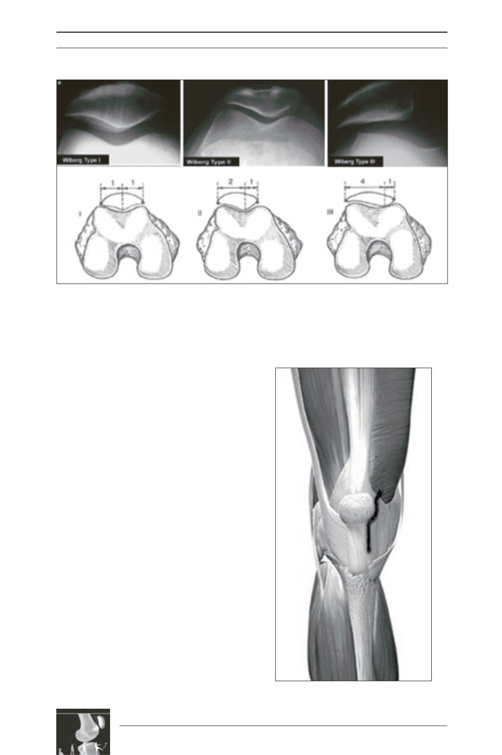
Each initial patellofemoral contourwas analysed
in order to rank the patellae according to the
Wiberg classification: types 1, 2, 3 (fig. 2).
The access chosen was the same for each patient
and performed by the same surgeon: mid-vastus
with preservation of the Hoffa (fig. 3).
At the lower part of the incision the patellar
tendon is carefully preserved up to its point of
termination on the TTA. If necessary for
exposure, subperiosteal infracentrimetric dis
placement of it was performed.
With the leg in extension, the patella is
systematically everted and dislocated.
The Hoffa is spared and does not restrict
exposure so much.
The arthroplasty is performed according to the
original “Lyon” technique. The tibial implant is
centred on the TTA. Femoral sectioning is
carried out with ligament balancing performed
in flexion then extension, taking care to avoid
any notching or femoral offset.
Fig. 2:
Wiberg type 1: medial and lateral surfaces concave and comparable in size
Wiberg type 2: medial surface smaller than the lateral surface with a concave appearance
Wiberg type 3: medial surface largely reduced
Fig. 2


