
D. Dejour, P.G. Ntagiopoulos
174
In 1998, Franck Remy and François Gougeon
published a study showing that the first 1987
classification of H. Dejour and G. Walch had
poor inter- and intra- observer reliability
especially for the type II. It was probably due
to the fact that the prominence or bump and the
shape of the facet and their asymmetry were
not taken into account in the three-type
classification [39, 40]. At that time they
observed the presence of the trochlear bump
and measured the trochlear depth in the
available X-rays, but did not correlate them to
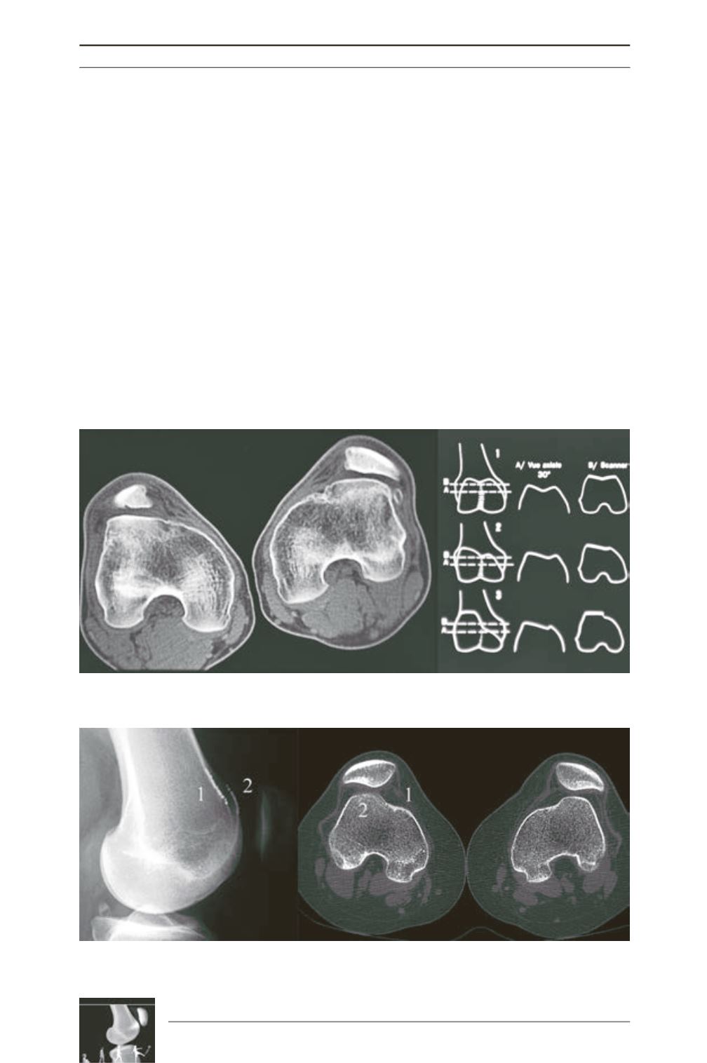
slice imaging (CT scan) (fig. 7). It was a three-
dimensional anomaly with a two-dimensional
imaging based only on X-rays. In 1998, David
Dejour and Bertrand Lecoultre did a study on
177 objective patellar dislocations comparing
the true sagittal view, the axial view and the CT
scan (fig. 8) [41]. Two more signs were added
to the definition of trochlear dysplasia: the
“supratrochlear spur” and the ‘double contour
sign’. The supratrochlear spur is the prominence
of the whole trochlea. It starts often proximal to
the cartilaginous part of the trochlea on the
lateral side (fig. 9). The double contour sign is
the osteochondral projection on the sagittal
view of the
hypoplastic medial facet
(fig. 8).
Using this three signs, they were able to publish
in 1998 a four grade classification which will
become in the future the most widely used
classification of trochlear dysplasia until now
[4, 41-43] (fig. 10):
Type A
involves mild
dysplasia where the trochlea
is shallower than
normal
with the characteristic crossing sign.
Fig. 7: (Original slide) The identification of trochlear bump and the measurement the trochlear
depth in X-rays that was not correlated to slice imaging at that time (left).
Fig. 8: The correlation of true lateral X-rays and slice imaging by D. Dejour and B. Lecoultre revealed the
presence of (1) the medial hypoplastic facet and the ‘double contour sign’, and (2) the supratrochlear spur.


